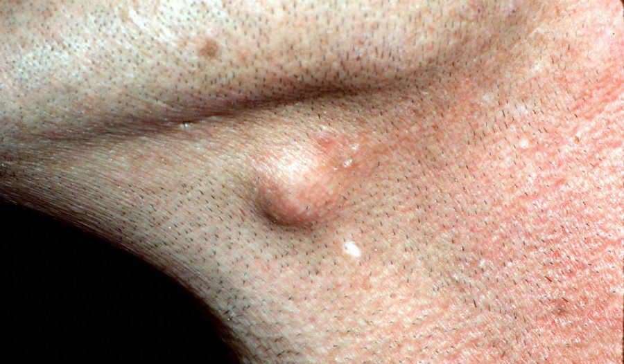
What is an epidermoid cyst?
An epidermoid cyst is a benign, slow-growing lump under the skin: a closed sac with a thin wall made of skin-type cells, filled with keratin, the protein that makes up the outer layer of the skin and hair (DermNet, BAD). It arises from the upper part of a hair follicle, which is why most cysts sit where there are follicles, but it can form almost anywhere (DermNet).
You will also hear it called an epidermal cyst, keratin cyst or infundibular cyst. The everyday name "sebaceous cyst" is a misnomer: the cyst has nothing to do with sebaceous (oil) glands and contains no sebum, only soft keratin (DermNet, BAD, MedlinePlus).
Skin cysts are very common, affecting at least 20% of adults (DermNet cysts). An epidermoid cyst is not cancer and cannot be passed to anyone else (NHS, BAD).
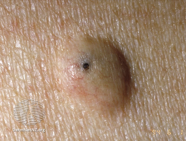
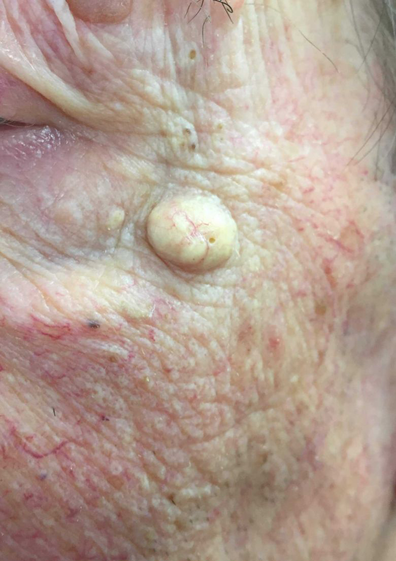
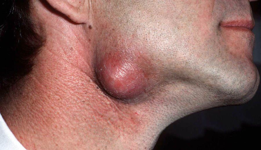
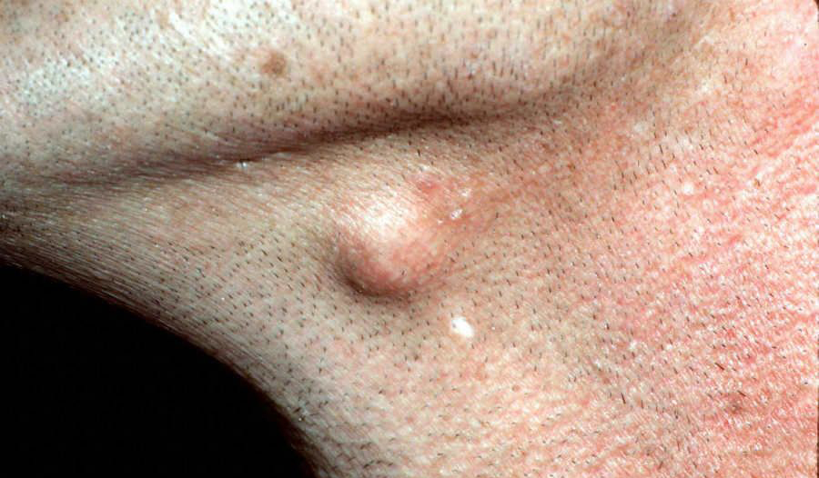
Epidermoid cysts: a dome-shaped nodule with the typical dark central punctum; a firm yellowish cyst on the cheek; an inflamed, red cyst on the neck; and a quiet skin-coloured cyst under the jawline.
Who gets epidermoid cysts?
They mostly affect adults, with a peak in the twenties and thirties (DermNet, BAD). Men are affected about twice as often as women (DermNet). People with acne get them more often (NHS), and so, occasionally, do people whose skin has been injured or operated on, because skin cells can be pushed below the surface and keep growing there (BAD, DermNet).
Multiple cysts appearing around puberty can be part of a rare inherited condition. Gardner syndrome, a form of familial adenomatous polyposis in which the bowel polyps almost always turn cancerous if untreated, produces multiple epidermoid cysts around puberty, alongside bony growths and lipomas (DermNet Gardner). A child or teenager with several cysts should therefore be assessed rather than reassured.
What causes an epidermoid cyst?
Most cysts start with a blocked hair follicle. The opening of the follicle is plugged, skin cells that would normally be shed at the surface keep multiplying inside, and the keratin they produce has nowhere to go. A sac forms and slowly fills (DermNet, MedlinePlus). On the palms and soles, and sometimes elsewhere after an injury, a cyst can form when a cut or a puncture carries surface skin cells into the deeper layer, where they build a sac around themselves: an epidermal inclusion cyst (DermNet, DermNet cysts).
Two things are not causes. Hygiene is not involved (BAD). And although the lump may look like a huge blackhead, it is a separate structure with its own wall, not a blocked pore you can clear (DermNet).
Cysts run in some families (MedlinePlus), and the inherited syndrome above accounts for a few cases of multiple cysts (DermNet).
Symptoms: what an epidermoid cyst looks and feels like
- A round lump under the skin, firm or slightly rubbery, fixed to the overlying skin but able to slide over the tissue beneath it (DermNet). It is usually painless (MedlinePlus).
- Size. Typically 1 to 3 cm, from smaller than a pea to several centimetres (DermNet, NHS). Cysts grow slowly and often stop at a certain size (MedlinePlus).
- Colour. Skin-coloured or yellowish (DermNet).
- The central punctum. Many cysts have a small dark dot or pore in the middle. This is the blocked follicle opening, the point where the sac connects to the surface (DermNet, BAD). Pressing the cyst makes it more obvious (DermNet). A punctum is a useful sign, because the main look-alikes, lipomas and pilar cysts, do not have one (DermNet lipoma, DermNet pilar).
- Contents. Thick, cheesy or toothpaste-like white-yellow keratin with a strong, unpleasant smell, which may leak from the punctum (DermNet, BAD, MedlinePlus).
- Location. Face, neck, chest, shoulders and upper back are the usual sites; the scrotum and vulva are also common, and there the cysts are often multiple (DermNet, BAD, NHS). In our online practice, a firm lump on the side of the neck or under the jaw is one of the commonest reasons people send a photograph.
An inflamed or infected cyst
Sometimes the thin wall tears and keratin spills into the surrounding tissue. The body treats it as a foreign substance, and within a day or two the cyst becomes red, swollen, warm and tender, and may enlarge quickly (DermNet, MedlinePlus). This inflammation is often sterile, and sterile pus may even be discharged; a true bacterial infection, usually with staphylococci, can follow and produces pus and sometimes fever (DermNet, DermNet cysts). On brown or black skin the redness is harder to see; warmth, swelling and tenderness are the better clues (NHS).
Is an epidermoid cyst dangerous?
No. An epidermoid cyst is a benign lesion that often stays unchanged for years, and some disappear by themselves (DermNet, NHS). The problems it can cause are:
- Rupture and inflammation, described above, which is the commonest complication (DermNet).
- Infection and abscess: a ruptured cyst can become secondarily infected and turn into a painful, pus-filled abscess (DermNet cysts, DermNet abscess, MedlinePlus).
- Recurrence after a cyst has ruptured or been incompletely removed, because any sac left behind refills (DermNet, BAD).
- Scarring from repeated inflammation or from surgery (DermNet, BAD).
- Cancer, very rarely. A squamous cell carcinoma has been reported arising inside an epidermoid cyst, but this is exceptional; it is one reason removed cysts are sent for microscopy (DermNet, DermNet SCC).
The more realistic danger is a wrong self-diagnosis. Some skin cancers form rounded nodules that look like a cyst. A nodular basal cell carcinoma is the usual culprit: it keeps enlarging, and eventually the surface breaks down, ulcerates and bleeds (DermNet cysts). Any new lump should be examined by a doctor rather than assumed to be a cyst (MedlinePlus).
Are cysts contagious?
No. There is no virus or bacterium behind an epidermoid cyst, only a sac of your own skin cells, so it cannot be passed on by touch, shared towels or sex (NHS, BAD). The one caveat is an infected cyst that is leaking pus: pus can carry bacteria, so cover a draining cyst and wash your hands, as you would with any wound.
Epidermoid cyst or something else?
A dermatologist tells these apart by feel (firm or soft, fixed or sliding), by the presence of a punctum, by where the lump is and how long it has been there, and sometimes by dermatoscopy or ultrasound. The common lumps that get confused with an epidermoid cyst:
| Lump | How it differs |
|---|---|
| Lipoma | A soft or rubbery, often lobulated fatty lump deeper under the skin, 1 to over 10 cm; the skin slides freely over it and there is no punctum. Grows slowly, rarely inflames (DermNet lipoma, NHS lipoma). |
| Pilar (trichilemmal) cyst | 90% are on the scalp; no punctum, thick wall that rarely ruptures, often several, and strongly familial (DermNet pilar, BAD). |
| Boil (furuncle) or abscess | Painful, hot, red lump with a soft centre that appears over days and fills with pus; may come with fever. Many boils settle by themselves, but a large, very painful or feverish one needs a doctor (NHS boils, NHS abscess). |
| Acne nodule or pseudocyst | No capsule, sits among blackheads, pimples and other acne lesions, and comes and goes with the acne (DermNet). |
| Milia | Tiny 1 to 2 mm pearly-white bumps, usually on the face and eyelids, often many (DermNet milium). |
| Steatocystoma | Multiple small, smooth, yellowish cysts on the chest, upper arms, armpits and neck, appearing in the late teens and twenties; filled with oily fluid rather than keratin (DermNet steatocystoma). |
| Dermoid cyst | Present from birth or early childhood, typically at the outer eyebrow, eyelid or along the neck; firm and dough-like (DermNet dermoid). |
| Dermatofibroma | A firm, often brownish button in the skin of the legs or arms that dimples inwards when squeezed from the sides (DermNet dermatofibroma). |
| Hidradenitis suppurativa | Recurrent painful boils and tunnels in the armpits, groin and under the breasts, usually several at once (DermNet hidradenitis). |
| Pilonidal sinus | A small pit or lump at the top of the cleft between the buttocks that becomes painful and discharges when infected (NHS pilonidal sinus). |
| Swollen lymph node | A rubbery lump at the side of the neck, under the jaw, in the armpit or groin, often with an infection nearby; usually settles within a couple of weeks (NHS swollen glands). |
| Nodular basal cell carcinoma | A pearly, shiny nodule that keeps growing, then ulcerates and bleeds; no punctum, no cheesy contents (DermNet cysts, DermNet basal cell carcinoma). |
When to see a doctor
See a GP or family doctor for any lump on your skin that you cannot explain, for a lump that is getting bigger, and for one that is hard and does not move, so that the diagnosis is confirmed (NHS, NHS abscess, BAD). Ask for an urgent appointment, or call 111 in the UK, if the lump:
- is sore, red or hot, or redness is spreading away from it;
- comes with feeling hot, cold or shivery, or a high temperature (NHS, NHS abscess).
Also see a doctor if the cyst keeps coming back, has ruptured, is on the face or genitals, snags on clothing or a comb, or simply bothers you (NHS, BAD). From clear photographs and your description of how the lump feels and how long it has been there, a dermatologist can usually judge whether it is likely to be an epidermoid cyst or one of its look-alikes; at iDerma we assess a lump online and advise whether it can be left, needs calming down or should be removed; the removal itself is done in person by a dermatologist or surgeon near you. You can book an online consultation and upload photographs; a lump that is hot and painful, or growing fast, should be seen in person without delay.
How an epidermoid cyst is diagnosed
In most cases the diagnosis is made by examination: a dermatologist judges size, consistency, mobility, the presence of a punctum and the state of the overlying skin (DermNet, BAD). No tests are needed for a typical cyst. Ultrasound is reserved for lumps whose depth or nature is uncertain (DermNet). If the cyst is infected, a swab may be taken to identify the bacteria (MedlinePlus). When a cyst is removed, the tissue is sent for microscopy to confirm the diagnosis and exclude the rare cancer (DermNet).
Treatment of an epidermoid cyst
The right treatment depends on whether the cyst is quiet, inflamed or infected, and on whether it bothers you.
A quiet cyst: leave it or remove it
A small cyst that causes no symptoms can safely be left alone (DermNet, NHS, BAD). Removal is reasonable when the cyst is large, catches on clothing or a comb, keeps becoming inflamed, or is in a visible place and you want it gone (NHS, BAD). The only treatment that removes a cyst for good is complete surgical excision of the whole sac under local anaesthetic, an injection that numbs the skin while you stay awake (DermNet, BAD, NHS local anaesthetic). The dermatologist numbs the skin, makes a small incision over the cyst, frees the intact sac from the surrounding tissue, lifts it out and closes the skin. If the wall is removed in one piece the cyst very rarely returns; if a fragment is left behind, it can refill (DermNet, BAD). Removal leaves a small scar (NHS, BAD). In the UK, removal of a cyst that causes no problems is usually not available on the NHS and is done privately (NHS).
An inflamed cyst
Inflammation after rupture is often sterile, so antibiotics alone frequently do little (DermNet pilar). Warm compresses several times a day can help the cyst settle or drain (MedlinePlus). A doctor may give an anti-inflammatory injection into the cyst to calm the swelling (MedlinePlus), or make a small incision to release the contents and relieve the pressure (DermNet, BAD). Drainage brings relief but leaves the sac in place, so the cyst can come back; the definitive excision is usually done later, once the inflammation has settled, because a ruptured, inflamed sac is difficult to remove in one piece (DermNet, BAD).
An infected cyst
If there is pus, spreading redness or fever, the cyst has become an abscess. It is drained, and antibiotics are prescribed when the infection is spreading or you are unwell (NHS, DermNet, DermNet abscess, NHS abscess). Removal of the sac can be arranged once the infection has settled (DermNet).
What not to do
Do not squeeze, pop or lance a cyst yourself. Pressure pushes keratin into the surrounding tissue and sets off exactly the inflammation you are trying to avoid; if the cyst is already infected, squeezing can spread the infection; and because the sac stays behind, the cyst comes back, often with a worse scar than a planned removal would leave (NHS, BAD, DermNet).
After removal: healing and scars
The wound is closed with stitches; keep it clean and dry as instructed, and avoid stretching or rubbing the area until the stitches are out. Every excision leaves a scar. Most scars fade and flatten over months, and continue to improve for up to two years (NHS scars). People who have formed a keloid before should say so beforehand, because the shoulders, chest and upper back, where cysts are common, are also the sites where raised scars are most likely (DermNet keloid). New cysts can form elsewhere; removal treats the cyst you have, not your tendency to make them (NHS, BAD).
Care and reducing flare-ups
You cannot prevent epidermoid cysts, and nothing about washing or skincare causes them (BAD). What helps:
- Leave a quiet cyst alone and do not squeeze it (NHS, BAD).
- Treat active acne, which is a risk factor for cysts (NHS); see our article on acne.
- Reduce friction on a cyst in an exposed spot, for example from a collar, bag strap or helmet strap; in our experience repeated rubbing often precedes a flare.
- Have recurrent or multiple cysts assessed, especially in a young person, because of the rare inherited syndromes (DermNet Gardner).
What to do now
- Note when the lump appeared, whether it has grown, whether it has ever been red or painful, and whether it has leaked.
- Photograph it in daylight, from the front and from the side, with a coin or ruler for scale.
- If it is hot, painful, spreading or you feel unwell, see a doctor today (NHS abscess). Otherwise book an online consultation with an iDerma dermatologist to confirm the diagnosis and discuss whether to leave it or remove it.
- Do not squeeze it in the meantime (NHS).