What is mycosis fungoides?
Mycosis fungoides is the most common form of cutaneous T-cell lymphoma, a cancer of T lymphocytes (a type of white blood cell) that begins in the skin. It is a form of non-Hodgkin lymphoma, not a skin cancer in the usual sense: the abnormal cells are immune cells living in the skin, not skin cells themselves (DermNet, Lymphoma Action, BAD). In most people it behaves like a long-term skin condition rather than an aggressive cancer. It usually develops over years, often stays in the skin, and most people are diagnosed at an early stage (DermNet, Lymphoma Action, Cutaneous Lymphoma Foundation).
The name is misleading. "Mycosis" suggests a fungus, but mycosis fungoides is not an infection; the name comes from the mushroom-like tumours that appear in advanced disease (NORD). It cannot be caught or passed on (BAD, Cutaneous Lymphoma Foundation).
- Rare: roughly 6 new cases per million people a year worldwide, about 450 diagnoses a year in the UK (DermNet, BAD).
- Common within its group: around half of all skin lymphomas and 60 to 70% of cutaneous T-cell lymphomas (DermNet, Lymphoma Action).
- Who: usually adults in their 50s and 60s, men about twice as often as women; children are affected rarely, and when they are, the pale (hypopigmented) form is the most common (DermNet, BAD).
- Outlook: the estimated 5-year survival is 87%; limited patch or plaque disease does not shorten life expectancy, although about one third of people with limited disease progress to a more advanced stage over time (DermNet).
What causes mycosis fungoides?
The cause is unknown. The leading theories are long-term, low-level stimulation of T cells in the skin, a genetic predisposition (for example particular HLA tissue types) and exposure to certain chemicals or medicines; clustering of cases in some regions and occasional cases within one family point to environmental triggers (DermNet, Cutaneous Lymphoma Foundation). Unlike some rarer skin lymphomas, there is currently no evidence that a virus causes mycosis fungoides (DermNet, DermNet Sézary).
Three things it is not:
- Not inherited. Families with more than one affected member exist but are very rare (BAD, Lymphoma Action).
- Not caused by sun exposure, unlike melanoma and other skin cancers; the patches in fact favour skin that is usually covered by clothing (Lymphoma Action, DermNet).
- Not caused by anything you did, and not contagious (Lymphoma Action, BAD).
For T-cell skin lymphomas as a whole, which include types that start later in life than classic mycosis fungoides, the factors linked to a higher risk are older age (two thirds of people are over 70 at diagnosis), male sex, African American ethnicity in US studies, a weakened immune system after an organ transplant or with HIV, and, for one specific subtype (adult T-cell leukaemia/lymphoma, not mycosis fungoides), infection with the HTLV-1 virus (Lymphoma Action CTCL).
Symptoms: what mycosis fungoides looks like at each stage
The rash usually develops slowly, often over many years, and can go through three skin stages. Different stages can be present on the body at the same time, and most people never progress beyond the first two (DermNet, Lymphoma Action CTCL, Cutaneous Lymphoma Foundation, BAD).
Itch, sometimes before any rash
Long-standing, generalised itching is the most common symptom and can start before anything is visible on the skin; around 80% of people with cutaneous T-cell lymphoma report itch. Pain in the affected areas is rare (DermNet, DermNet CTCL, BAD).
Patch stage (early mycosis fungoides)
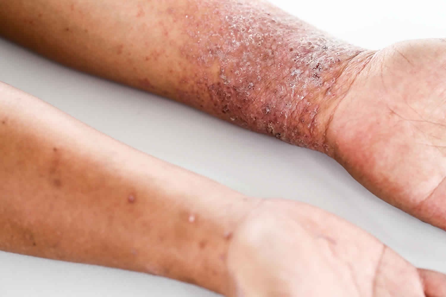
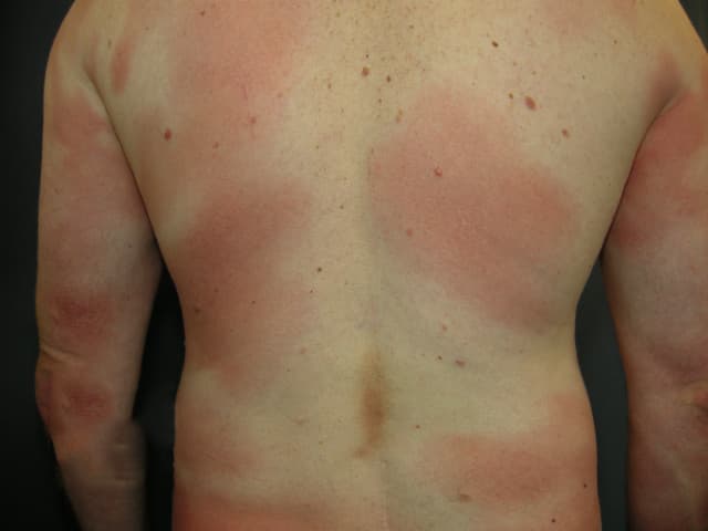
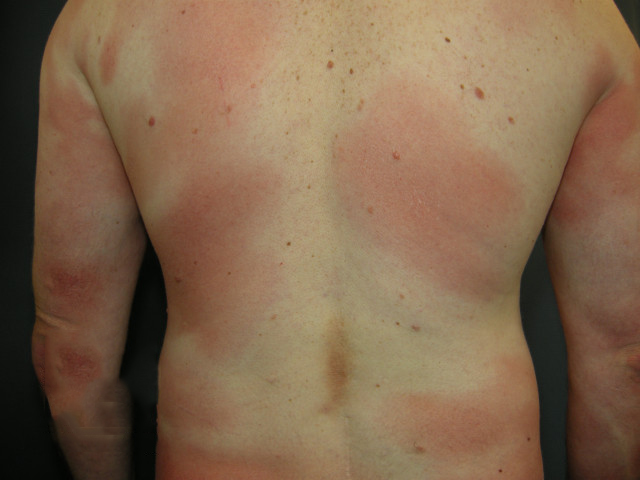
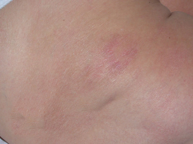
- Flat, irregular, poorly defined red or pink patches of varying size, with fine scale and often thin, wrinkled skin on the surface (DermNet).
- They favour skin that the sun does not reach: the buttocks, lower trunk, thighs and, in women, the breasts (DermNet, Lymphoma Action).
- Patches are often dry and may itch, but many cause no symptoms at all; they can stay the same for years, slowly enlarge, or disappear on their own for a while (DermNet, Lymphoma Action CTCL).
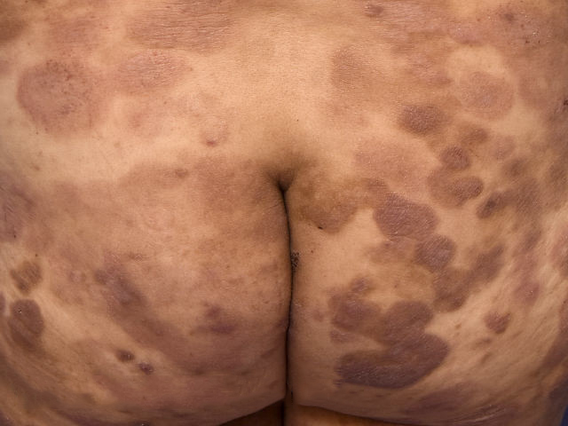
- On brown or black skin, patches are more often lighter (hypopigmented) or darker than the surrounding skin rather than red; in children the pale form is the usual presentation (DermNet, Lymphoma Action, Cutaneous Lymphoma Foundation).
- Because the patches look like eczema or psoriasis, this stage is frequently treated as one of those conditions for years before anyone suspects lymphoma (BAD, Lymphoma Action, Cutaneous Lymphoma Foundation).
Plaque stage
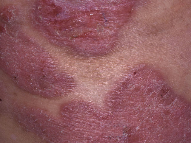
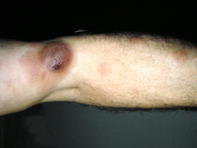
- Thicker, raised, well-defined areas, often ring-shaped or arc-shaped, red, violet or brown, sometimes scaly; they can grow quite large and clear in the centre (DermNet).
- Plaques can grow out of existing patches or appear on new skin; early on they cover less than a tenth of the skin surface (DermNet).
- They are usually itchy and can break down (ulcerate); hair may be lost in affected areas (Lymphoma Action CTCL).
Tumour stage
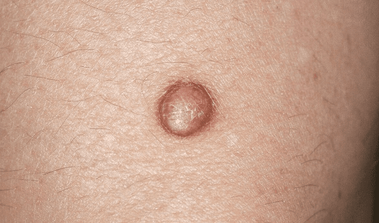
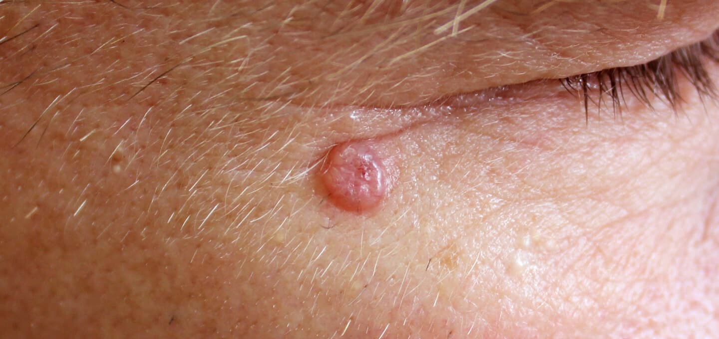
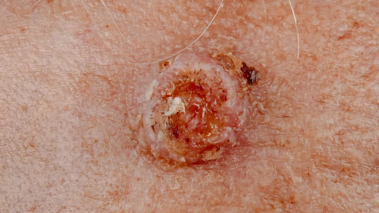
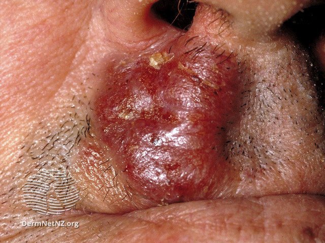
- Lumps of 1 cm or more, deep red to violet, often with a shiny surface, usually growing out of plaques and occasionally appearing without an earlier plaque (DermNet, NCI, NORD).
- Tumours may ulcerate, weep, become painful and get infected (Lymphoma Action CTCL, NCI).
- Only a small proportion of people ever reach this stage, and tumours are rarely the first sign (Lymphoma Action CTCL, Cutaneous Lymphoma Foundation).
Erythroderma and spread beyond the skin
Rarely, the skin becomes red, thickened, scaly and intensely itchy over most of the body (erythroderma), the palms and soles may thicken and crack, and lymph nodes in the neck, armpits or groin may swell. When large numbers of abnormal cells are also circulating in the blood, the condition is called Sézary syndrome, a rarer, high-grade form of cutaneous T-cell lymphoma that is staged together with mycosis fungoides; it usually arises on its own and is only rarely preceded by mycosis fungoides, and whether it is a separate disease or an advanced form is still debated (Lymphoma Action CTCL, DermNet Sézary, NCI). Spread to lymph nodes or internal organs occurs in about 10% of people with cutaneous T-cell lymphoma and almost never happens without obvious worsening in the skin first (DermNet CTCL, Cutaneous Lymphoma Foundation).
Less typical forms
Several variants follow the same slow course but look different. Folliculotropic mycosis fungoides, the most common of them, affects hair follicles, usually on the head and neck, with itchy grouped bumps that can resemble acne or keratosis pilaris, blocked pores or small cysts, and patchy hair loss, often in the eyebrows (DermNet, Lymphoma Action CTCL). Mycosis fungoides can also mimic pigmented purpura (capillaritis), granuloma annulare, vitiligo or psoriasis, or present as a single scaly patch or warty plaque on a limb (pagetoid reticulosis), or as loose folds of skin in the armpits and groin (granulomatous slack skin) (DermNet, Lymphoma Action CTCL).
Is mycosis fungoides dangerous?
For most people it is no more dangerous than a long-term skin condition. Mycosis fungoides is a cancer by definition, but it typically follows a slow course over years, and limited patch or plaque disease does not alter life expectancy (DermNet, BAD). The estimated 5-year survival is 87%, early-stage disease has been estimated to last around 18 years on average, and many people live a normal life span (DermNet, BAD, Cutaneous Lymphoma Foundation). The outlook depends mainly on age, how the skin is affected (patches and plaques versus tumours or erythroderma) and whether lymph nodes or internal organs are involved; the pale and poikilodermatous variants tend to progress less, while folliculotropic, pustular and granulomatous forms behave more aggressively (DermNet).
Two complications matter in practice: ulcerated plaques or tumours can become infected, and the uncertainty of a lifelong relapsing condition commonly brings anxiety and low mood (DermNet, DermNet CTCL).
The four stages doctors use
Once the diagnosis is confirmed, the disease is staged by how much skin is affected and whether the lymph nodes, blood or internal organs are involved. Most people are diagnosed at stage 1 or 2A, which doctors call early-stage disease (Lymphoma Action CTCL, NCI).
| Stage | What it means |
|---|---|
| 1A | Patches or plaques only, covering less than a tenth of the skin surface (NCI, Lymphoma Action CTCL). |
| 1B | Patches or plaques covering a tenth or more of the skin surface; a tenth is roughly the area of both arms (Lymphoma Action CTCL). |
| 2A | Patches or plaques with enlarged lymph nodes that do not contain lymphoma cells (NCI). |
| 2B | One or more skin tumours of 1 cm or larger; lymph nodes may or may not be enlarged but do not contain lymphoma cells (NCI, Lymphoma Action CTCL). |
| 3 | Redness over four fifths or more of the skin (erythroderma), with no, few or a moderate number of abnormal cells in the blood (Lymphoma Action CTCL, NCI). |
| 4 | Numerous abnormal cells in the blood (the picture doctors call Sézary syndrome), lymphoma in the lymph nodes, or spread to other organs such as the liver or spleen (NCI, Lymphoma Action CTCL). |
Conditions that look like mycosis fungoides
Most dry, red, scaly patches are not lymphoma. All of the conditions below are far more common, and several of them improve with the same steroid creams used for early mycosis fungoides, which is one reason the diagnosis is often delayed (Lymphoma Action). None of them can be told apart from mycosis fungoides with certainty by eye; that needs a skin biopsy (Lymphoma Action, DermNet).
| Condition | How it typically differs |
|---|---|
| Atopic eczema | Usually starts in childhood, favours the elbow and knee creases and hands, flares and settles, and runs with asthma or hay fever (DermNet atopic dermatitis, NHS eczema). |
| Psoriasis | Well-defined plaques with thick silvery scale on the elbows, knees, scalp and lower back; nail pitting; often a family history (DermNet psoriasis, NHS psoriasis). |
| Discoid (nummular) eczema | Scattered coin-shaped plaques of 1 to 3 cm, mostly on the legs, either oozing and blistered or dry and scaly (DermNet discoid eczema). |
| Chronic superficial scaly dermatitis (parapsoriasis) | Finger-shaped pink scaly patches on the trunk and limbs, often worse in winter; the large-plaque form is considered a possible precursor, and 7.5 to 14% of large plaques progress to mycosis fungoides, so these are monitored (DermNet CSSD). |
| Ringworm of the body (tinea corporis) | A circular red patch with a raised scaly leading edge that spreads outwards into a ring and clears with antifungal treatment; often caught from pets or family members (DermNet tinea corporis, NHS ringworm). |
| Pityriasis versicolor | Fine-scaled pink, pale brown or pale patches on the chest and upper back that do not tan; a yeast overgrowth, more common in hot weather (DermNet pityriasis versicolor, NHS pityriasis versicolor). |
| Pityriasis rosea | A single oval "herald patch" followed by smaller oval patches in a fir-tree pattern on the trunk; clears by itself in 6 to 10 weeks (DermNet pityriasis rosea, NHS pityriasis rosea). |
| Vitiligo and pityriasis alba | Look-alikes of the pale form: vitiligo patches gradually turn completely white; pityriasis alba gives finely scaly pale patches, mainly in children with dry or eczema-prone skin (NHS vitiligo, DermNet pityriasis alba). |
| Contact dermatitis | A reaction where an irritant or allergen touched the skin, either from repeated exposure to soaps, water or friction or from a true allergy that can spread beyond the contact area; patch tests identify the allergen (DermNet contact dermatitis). |
| Lichen simplex (neurodermatitis) | One or a few very itchy, thickened, leathery plaques caused by repeated rubbing or scratching (DermNet lichen simplex). |
| Scabies | Intense itch with a rash in a characteristic distribution, from a mite that burrows into the skin; highly contagious, so household contacts itch too (DermNet scabies). |
| Drug rashes | A widespread measles-like rash, or rarely erythroderma, after starting a new medicine (DermNet drug eruptions, DermNet erythroderma). |
| Pityriasis lichenoides and lymphomatoid papulosis | Crops of small scaly or crusted spots that heal and recur; pityriasis lichenoides is confused with the pale form of mycosis fungoides, and lymphomatoid papulosis is itself a harmless relative of T-cell skin lymphoma that needs a specialist to interpret (DermNet pityriasis lichenoides, DermNet lymphomatoid papulosis, Lymphoma Action CTCL). |
When to see a doctor
- A dry, scaly or discoloured patch that has not cleared after a 4 to 6 week course of a prescribed steroid cream, or that keeps coming back in the same place, particularly on the buttocks, trunk or thighs (Lymphoma Action, Lymphoma Action treatments, BAD).
- A rash diagnosed as eczema or psoriasis that does not behave as expected, or itching that has gone on for weeks with no obvious cause (DermNet, Lymphoma Action, NHS itchy skin).
- Patches that are thickening, forming lumps, breaking down or weeping (Lymphoma Action CTCL).
- Swollen glands in the neck, armpits or groin that are getting bigger, feel hard, do not move when pressed or have not gone down within a week, especially with night sweats, fevers or unexplained weight loss (NHS swollen glands, NHS non-Hodgkin lymphoma symptoms).
- Redness, scaling and itch spreading over most of the body: this is potentially serious and needs prompt medical assessment, whatever the cause (DermNet erythroderma).
An online dermatology review is a reasonable first step for a persistent patch: send sharp photographs in daylight of each affected area, note how long each patch has been there and what creams you have used, and an iDerma dermatologist can advise whether a biopsy is needed and how to arrange a referral. Confirming or excluding mycosis fungoides always requires an in-person biopsy, so a remote consultation can guide you but cannot replace it.
How mycosis fungoides is diagnosed
Diagnosis rests on matching what the doctor sees with what the pathologist finds in the skin, and it is often slow. The changes under the microscope are subtle in the early stages, so biopsies frequently have to be repeated over months or years; on average it takes about three years to confirm a skin lymphoma, and some people have seven or eight biopsies (DermNet, BAD, Lymphoma Action, Lymphoma Action CTCL). The wait is frustrating, but early treatment is not critical for mycosis fungoides, and for early-stage disease a delay in diagnosis does not usually change the overall outlook; that is a reason not to panic, not a reason to leave a persistent patch unchecked (Lymphoma Action, Lymphoma Action CTCL).
- History and examination: how the rash developed, whether it itches and comes and goes, which treatments have been tried; the whole skin is examined, lymph nodes are felt, and the skin may be photographed to track change (Lymphoma Action, NCI). Dermoscopy helps separate mycosis fungoides from inflammatory rashes (DermNet).
- Skin biopsy: under local anaesthetic a small full-thickness sample, usually a punch of 3 to 4 mm, is removed and examined for the typical infiltrate of abnormal CD4+ T cells; special stains, flow cytometry and T-cell receptor gene tests can show whether the cells are a single clone (DermNet, DermNet skin biopsy, NCI). Results can take 4 to 6 weeks (Lymphoma Action).
- Blood tests: a full blood count, which is normal in most people with early disease, a count of abnormal (Sézary) cells, and tests for infections such as HIV (BAD, NCI, Lymphoma Action).
- Scans and further biopsies: a CT or PET-CT scan, a lymph node biopsy if a node is enlarged, and rarely a bone marrow biopsy are used for staging in more advanced disease (DermNet, Lymphoma Action, NCI).
Because the condition is rare, the diagnosis should be confirmed by a pathologist experienced in cutaneous lymphoma, and people are looked after by a multidisciplinary team that can include a dermatologist, a haematologist or clinical oncologist and specialist nurses (Cutaneous Lymphoma Foundation, Lymphoma Action, BAD).
Treatment
Mycosis fungoides is treatable but not curable. The aim is to control symptoms, clear the skin for as long as possible and protect quality of life with as few side effects as possible; no treatment has been shown to lengthen survival, and relapse after treatment is common (DermNet, BAD, Lymphoma Action treatments). Treatment is chosen according to stage, symptoms, age, general health and personal preference, and many people have several different treatments over their lifetime, with periods of no treatment in between (BAD, Lymphoma Action treatments).
Active monitoring
Early, limited disease that is not troublesome may simply be watched at regular clinic visits, because the potential benefit of treating it is smaller than the risk of side effects. Good skin care with regular moisturisers, including prescribed urea-based emollients for very dry skin, may be all that is needed at stage 1 (DermNet, Lymphoma Action, Lymphoma Action CTCL).
Skin-directed treatments
Most people only ever need treatments applied to the skin (BAD):
- Steroid creams and ointments. Potent prescription steroids can clear patches after 4 to 6 weeks and are often the only treatment needed in early disease; a dermatology team will explain how to use them safely (BAD, Lymphoma Action treatments).
- A chemotherapy gel applied to the affected skin, used for over 50 years; it can take up to a year to clear patches and causes skin irritation in up to 4 in 10 people (BAD, Lymphoma Action treatments).
- Phototherapy (light treatment). Narrowband UVB, or UVA combined with a light-sensitising medicine (PUVA), given two or three times a week in a walk-in cabinet for 15 to 30 sessions; remissions after PUVA can last up to 5 years. Because UV light raises the long-term risk of skin cancer, there is a lifetime limit on sessions (Lymphoma Action treatments, DermNet UVB, DermNet PUVA).
- Radiotherapy. Skin lymphoma is usually very sensitive to radiotherapy: low-dose treatment can clear individual plaques or tumours, and whole-skin electron beam treatment, given at specialist centres, treats the entire skin surface when other treatments have failed (BAD, Lymphoma Action treatments).
- Other creams and gels used for limited areas when steroids have not helped include vitamin A-related (retinoid) preparations and immune-modifying creams and ointments, prescribed by a dermatologist outside the product's licence ('off-label') (Lymphoma Action treatments).
Whole-body (systemic) treatments
If skin-directed treatment no longer controls the disease, or it is advanced at diagnosis, a specialist skin lymphoma team may add treatments that work throughout the body (BAD, Lymphoma Action CTCL):
- Injected immune-stimulating treatment that encourages the immune system to attack the lymphoma cells and stops them dividing, often combined with PUVA (BAD, Lymphoma Action treatments).
- Retinoid (vitamin A-related) capsules that disrupt the growth and division of lymphoma cells, with regular blood tests to watch blood fats and thyroid levels (BAD, Lymphoma Action treatments).
- Once-weekly tablets that slow down the production of new cells (BAD).
- Antibody treatments that bind to proteins on the lymphoma cells, used when the disease has returned or not responded to earlier systemic treatment (BAD, NCI).
- Extracorporeal photopheresis, in which some blood cells are taken out, treated with UVA light and returned, mainly for erythroderma (BAD, NCI, Lymphoma Action CTCL).
- Conventional chemotherapy, used sparingly because mycosis fungoides responds poorly to it and tends to return quickly (DermNet, Cutaneous Lymphoma Foundation).
- A donor stem cell transplant, considered for younger, fit people with advanced disease that has not responded to other treatment (BAD, Lymphoma Action CTCL).
When symptoms flare you do not automatically need a stronger treatment than before; the usual approach is the lowest-intensity option that controls the skin (Lymphoma Action CTCL). People with mycosis fungoides may be invited to join clinical trials, including trials of newer immune-checkpoint treatments (NCI, Cutaneous Lymphoma Foundation).
Living with mycosis fungoides
- Moisturise daily with an unperfumed emollient to reduce dryness, irritation and itch; emollients soothe but do not treat the lymphoma itself (BAD, Lymphoma Action treatments).
- For itch: pat rather than scratch, keep showers lukewarm and short, use a sensitive-skin laundry product, wear loose cotton, and ask a pharmacist or your team about antihistamines or cooling creams (NHS itchy skin).
- Protect treated skin from the sun during and after phototherapy or radiotherapy, and wear UV-protective sunglasses on PUVA days (Lymphoma Action treatments).
- No special diet is needed, and there are usually no restrictions on daily activities (BAD).
- Watch for change: photograph your patches regularly and report thickening, new lumps, ulcers, swollen glands, fevers or weight loss (Lymphoma Action CTCL, Cutaneous Lymphoma Foundation).
- Ask for support. Anxiety and low mood are common with a visible, lifelong condition; specialist nurses and patient organisations such as Lymphoma Action and the Cutaneous Lymphoma Foundation offer information and peer support (DermNet CTCL, BAD).
How iDerma can help
If you have a scaly patch that has not cleared, or an "eczema" that never quite behaves like eczema, you can book an online consultation. A dermatologist reviews your photographs and history, tells you whether the pattern raises concern, and recommends the next step, which for suspected mycosis fungoides is a referral for a skin biopsy. If you already have a diagnosis, an online review is useful for questions about skin care, itch and recognising a flare between hospital appointments.
Photo credit: the sixth gallery image is reproduced from DermNet NZ.