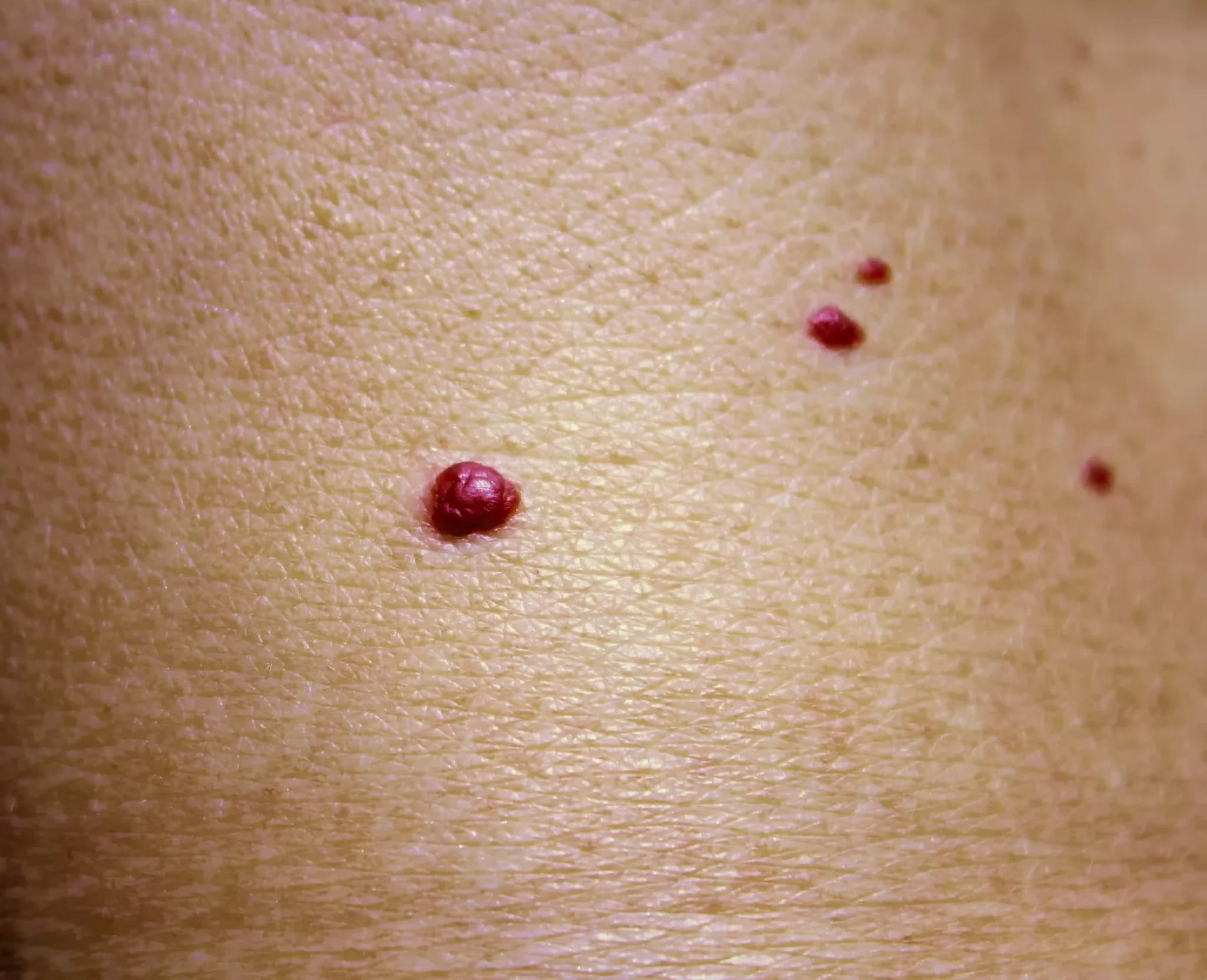
What is a cherry angioma?
A cherry angioma is a small, bright red, smooth bump made of tiny blood vessels. It is the most common type of angioma, a benign (non-cancerous) growth that forms when the cells lining a blood vessel multiply (DermNet). Other names are Campbell de Morgan spot, senile angioma, cherry haemangioma and, in everyday language, “red mole” or “blood spot” (DermNet).
Cherry angiomas are harmless. They do not turn into cancer, they are not infectious and they do not need treatment (DermNet, MedlinePlus, Cleveland Clinic). The only practical problem is bleeding when one is scratched or knocked, and the only medical question is making sure a red or black spot really is an angioma and not one of the few look-alikes that matter (DermNet).
Strictly, an angioma is any benign growth of vessels in the skin, and a haemangioma (American spelling “hemangioma”) is one made of blood vessels rather than lymph vessels (DermNet, Cleveland Clinic). Angiomas can appear in babies (infantile haemangioma, the “strawberry mark”) or later in life, and the cherry angioma is the common adult form (DermNet).
Who gets cherry angiomas?
- Almost everyone eventually. The number of spots increases markedly from about the age of 40, and an estimated 75% of people over 75 have them (DermNet). About half of adults have at least one after the age of 30 (Cleveland Clinic).
- They are not only a sign of age: about 5% of adolescents have them too (DermNet).
- Men and women, and people of every skin colour, are affected equally; the spots are simply more visible on white skin than on skin of colour (DermNet).
- They often run in families (DermNet, MedlinePlus).
- Pregnancy can trigger new angiomas, and some of those shrink or disappear after delivery (Cleveland Clinic, DermNet).
What causes cherry angiomas?
The cause is not known. Genetic analysis has found that many cherry angiomas carry small acquired mutations in two genes (GNAQ and GNA11) in the cells of the spot itself; these are not inherited and are also seen in other vascular growths (DermNet). What makes a particular person form many of them is less clear. The factors that are linked to new angiomas are (Cleveland Clinic, DermNet):
- ageing, the strongest factor by far;
- pregnancy hormones;
- a family tendency;
- exposure to certain chemicals (bromide compounds and an industrial solvent) and one topical medical treatment (Cleveland Clinic).
Two popular explanations do not hold up. Sun exposure is not among the causes any of these sources lists, and the commonest site is the trunk, which is usually covered (MedlinePlus, Cleveland Clinic). Liver disease is not the explanation either: the red spots associated with liver disease are spider telangiectases (spider naevi), a different lesion with a central dot and radiating fine vessels, as DermNet describes on its spider telangiectasis page (DermNet).
What does a cherry angioma look like?
- Size and shape: a round spot from a pinhead to about 1 cm across, smooth, either flat or slightly raised (DermNet, MedlinePlus). Cleveland Clinic gives 1–5 mm as the usual range, sometimes with a pale halo around the spot (Cleveland Clinic).
- Colour: cherry-red, sometimes purple or blue. A spot can turn black suddenly if the blood inside it clots (see below) (DermNet, PCDS).
- Texture: soft to firm and partly compressible; pressing does not make it disappear the way it does with a spider naevus (PCDS, DermNet). If it feels scaly or warty it is more likely an angiokeratoma (PCDS).
- Number: usually several to many, scattered over the body, and the count rises with age (DermNet, PCDS).
- Sites: most often the trunk (chest, abdomen, back), then the arms and legs (MedlinePlus, Cleveland Clinic). They can appear almost anywhere but are rare on the hands, feet and mucous membranes (DermNet).
- Symptoms: none. A cherry angioma does not hurt or itch (DermNet, Cleveland Clinic). It can bleed if scratched, shaved or rubbed by clothing, because it is made of blood vessels (Cleveland Clinic, PCDS).
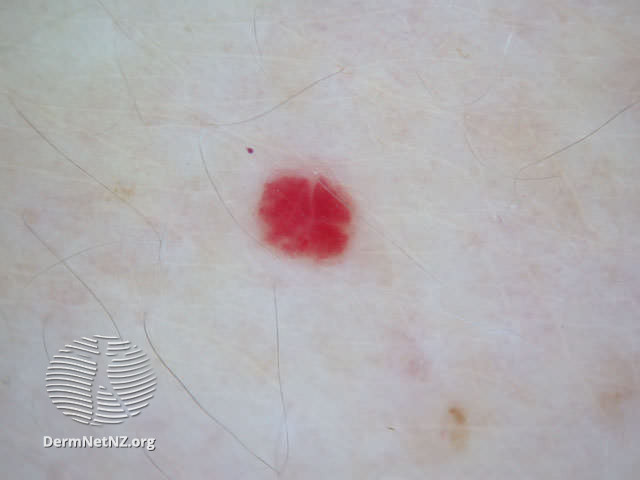
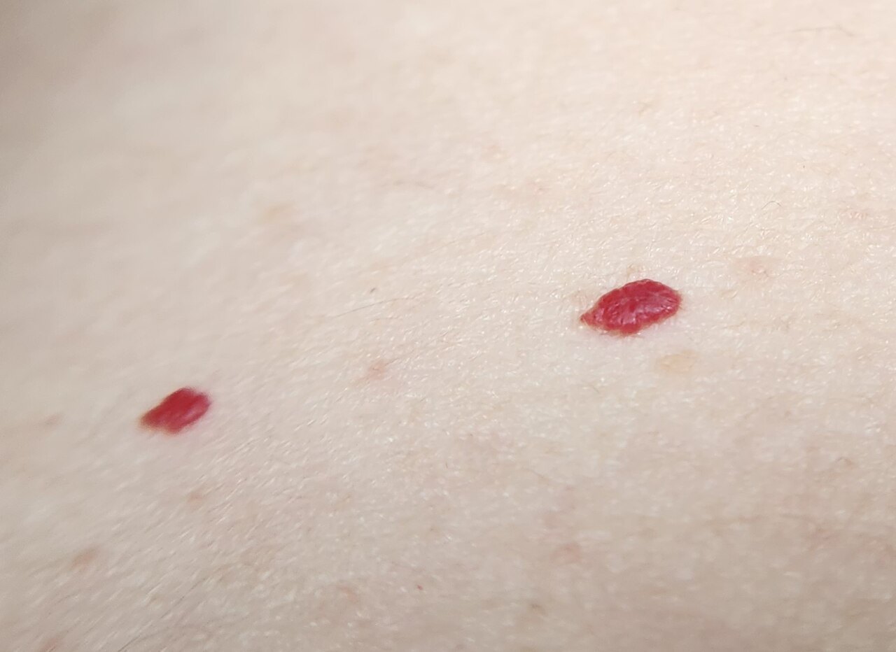
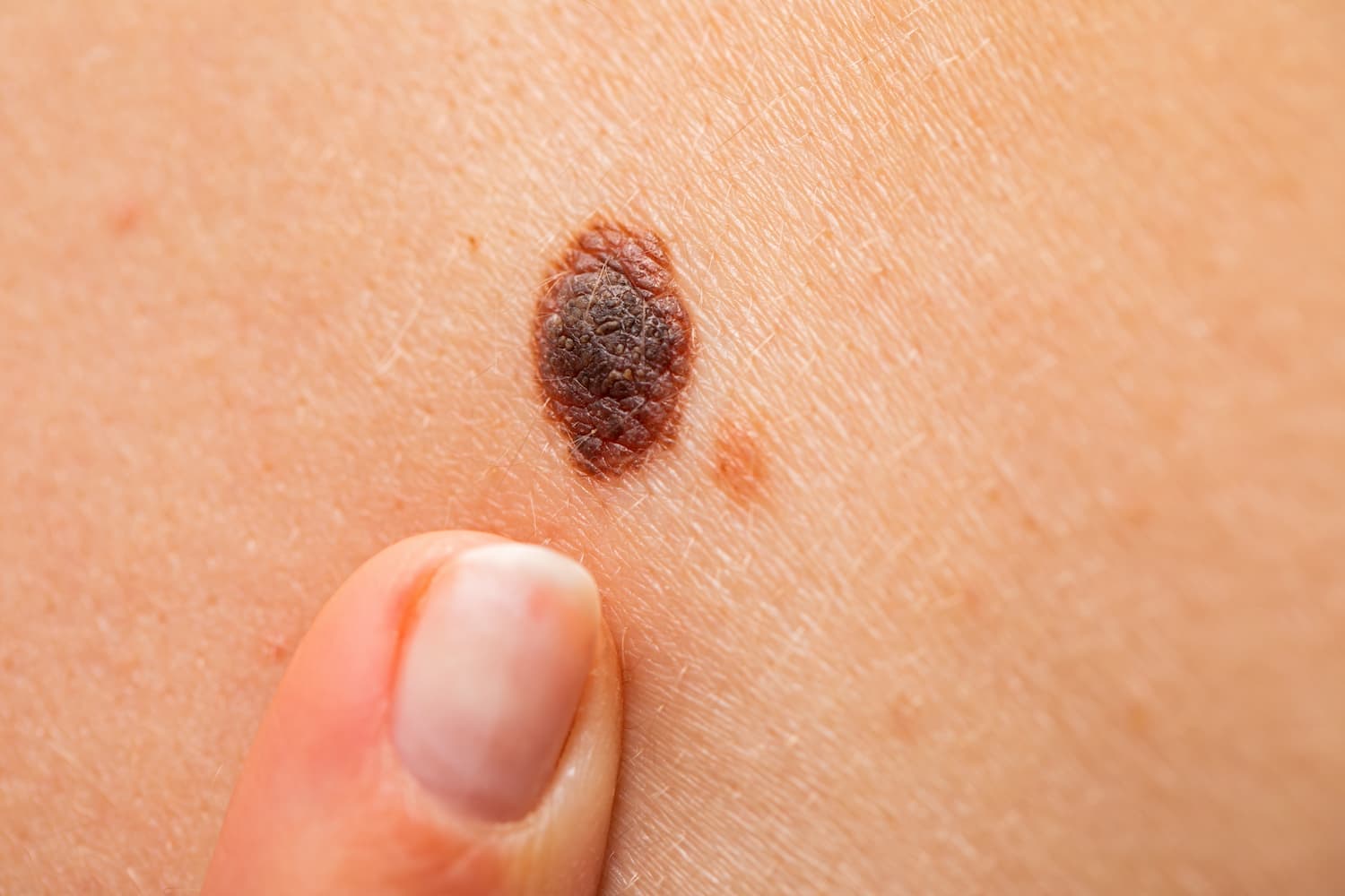
Left: a single cherry angioma. Right: two cherry angiomas close up, bright red, smooth and sharply outlined.
A black cherry angioma. When the blood inside an angioma clots (thrombosis), the spot turns purple-black or black overnight. A thrombosed angioma is harmless, but by eye alone it cannot be told apart from a nodular melanoma. Under a dermatoscope the red or purple colour usually shows through again; if any doubt remains, the spot is removed and examined under the microscope (DermNet, PCDS). A spot that has suddenly turned black should always be shown to a dermatologist in person.
Why am I suddenly getting many cherry angiomas?
For most people the answer is age: new spots keep appearing from the forties onwards, and it is normal to get a few new ones every year (DermNet). Pregnancy is the other common reason, and some of those spots fade after the birth (Cleveland Clinic). A true “eruption”, dozens of new angiomas within weeks, is uncommon. DermNet notes that eruptive cherry angiomas have rarely been linked with an internal cancer, and Cleveland Clinic lists exposure to bromide compounds, an industrial solvent and one topical medical treatment among the triggers (DermNet, Cleveland Clinic). A sudden crop of many new spots, especially together with weight loss, tiredness, night sweats or fever, is a reason to see a doctor, not because the spots are dangerous but because the trigger should be looked for.
Is a cherry angioma dangerous?
No. A cherry angioma is benign and stays benign; it cannot become a skin cancer (DermNet, MedlinePlus, Cleveland Clinic). The risk lies in the opposite direction: a red or pink lump that is not an angioma can be dismissed as one. Amelanotic melanoma, the form of melanoma with little or no pigment, makes up roughly 2–20% of all melanomas and is often red or pink; it may lack the classic ABCDE warning signs, which is why dermatologists add the “3 Rs”: Red, Raised, Recent change (DermNet). Nodular melanoma presents as a firm, dome-shaped lump that grows over weeks to months, is often larger than 6 mm and in one third of cases has no brown or black pigment at all (DermNet). A cherry angioma, in contrast, typically stays the same size and colour for years.
So the rule is simple: a red spot that looks like your other red spots and has not changed is almost certainly a cherry angioma. A single spot that is new, growing, changing colour, bleeding without being knocked, or that looks different from the rest needs to be examined (NHS, HSE).
Cherry angioma or something else? The look-alikes
| Lesion | How it differs from a cherry angioma |
|---|---|
| Spider telangiectasis (spider naevus) | A central red dot with fine red lines radiating from it; it blanches completely when pressed and refills at once. Common on the face, neck and upper chest. Several at once are linked with pregnancy, the contraceptive pill and liver disease (DermNet). |
| Angiokeratoma | A blue-red to purple papule, seldom more than 5 mm, with a rough, warty or scaly surface; becomes more warty with time; can bleed or clot and become painful (DermNet, PCDS). |
| Pyogenic granuloma | A red, fleshy nodule 5–10 mm that appears and grows over a few weeks, often on a finger or the face, after minor injury or in pregnancy; bleeds very easily and may crust (DermNet). |
| Venous lake | A soft, squashable blue or purple spot 2–10 mm, usually on the lower lip or an earlobe of an older person; it empties when pressed (DermNet). |
| Amelanotic or nodular melanoma | A new or changing pink, red, skin-coloured or black lump that grows over weeks to months, may bleed or itch, and often lacks the ABCDE signs. Remember the 3 Rs: Red, Raised, Recent change (DermNet). |
| Nodular basal cell carcinoma | A shiny or pearly nodule with fine visible vessels that keeps growing over months and may ulcerate or crust; usually on sun-exposed skin. A growth that bleeds, crusts or scabs for more than 4 weeks should be checked (DermNet, NHS). |
| Kaposi sarcoma | Red to purple patches, plaques or nodules, often starting on the lower legs, usually in people with weakened immunity, though a slow 'classic' form occurs in older men; linked to a herpesvirus (HHV8) (DermNet). |
| Bacillary angiomatosis | Red-purple papules and nodules that bleed easily, from a bacterial infection in people with weakened immunity, often with fever (DermNet). |
| Glomus tumour | A single reddish-blue 1–2 cm nodule on or beside a nail bed that is very painful, especially with cold or pressure (DermNet). |
| Dermatofibroma | A firm brown-to-pink nodule, usually on a leg, that dimples inwards when pinched (DermNet). |
| Infantile haemangioma | The “strawberry mark” of babies: appears in the first weeks of life, grows fast for about 3 months, then slowly shrinks over several years (DermNet). |
| Ordinary moles | Usually brown, round or oval with a smooth edge; harmless moles usually do not change (NHS). |
Two more things cause confusion. A tiny fresh haematoma or blood blister after a knock is dark red to black and disappears within weeks, whereas a cherry angioma stays. And a seborrhoeic keratosis or a skin tag (soft fibroma) can be skin-coloured or pale, but neither has the glassy red colour of a vascular spot (DermNet, NHS).
When to see a doctor
Cherry angiomas themselves never need an appointment. See a dermatologist, or your GP first in the UK and Ireland, if (DermNet, NHS, HSE, Cleveland Clinic):
- a spot has changed size, shape or colour, or has an uneven edge;
- a spot has turned black, or a single dark spot has appeared on its own;
- a spot bleeds without being knocked, or keeps bleeding, crusting or scabbing for more than 4 weeks;
- a red lump is new and growing (Red, Raised, Recent change);
- dozens of new spots have appeared within a few weeks;
- a spot is constantly rubbed by a bra strap, belt, collar or razor and bleeds;
- you are not sure what the spot is, or you want it removed.
How a cherry angioma is diagnosed
In most cases a dermatologist recognises a cherry angioma by eye and no test is needed (DermNet, MedlinePlus). The diagnosis can be confirmed with dermatoscopy, a hand-held magnifier with its own light: a cherry angioma shows a characteristic “lacunar” or red-clod pattern, round red or purple pools separated by pale lines, and in most spots no individual vessels are seen (DermNet, PCDS). Any other colour, such as brown, or vessels running through the lesion are red flags that put melanoma back on the list (PCDS). A 2026 study of 380 angiomas in 99 adults found the classic pools and lines in the larger spots, while the smallest spots mainly showed fine looped vessels, so tiny early angiomas can look less typical under the dermatoscope (González-Valdés 2026).
A biopsy is reserved for the uncertain case, typically a black or rapidly growing lump, and shows a cluster of small dilated vessels in the upper layer of the skin (DermNet). Typical red spots can be assessed from good photographs in an online consultation; a black, growing or bleeding lump needs to be looked at under a dermatoscope in person.
Do cherry angiomas need treatment?
No. Because they are harmless, treatment is optional and is chosen for one of three reasons (DermNet, MedlinePlus):
- the spot is in a visible place and bothers you (face, neck, décolletage);
- it is repeatedly knocked or rubbed and bleeds;
- the dermatologist wants to rule out a melanoma, in which case the spot is removed in a way that allows it to be examined under the microscope (DermNet).
As with harmless moles, cosmetic removal is not normally provided by the NHS or the HSE; you pay a private clinic (NHS, HSE). The removal itself is done in person by a dermatologist or surgeon near you; an online consultation can tell you whether removal makes sense and which method would suit you.
Removal methods
- Electrosurgery (electrocautery, electrodesiccation): the spot is destroyed with a fine electric needle under local anaesthetic; electrocoagulation also seals the vessels, which is why it suits vascular lesions (DermNet, MedlinePlus, Cleveland Clinic).
- Vascular laser: laser light absorbed by haemoglobin, the red pigment in blood, heats and closes the vessels. The pulsed dye laser is the usual choice for vascular lesions; it leaves bruising for 1–2 weeks and sometimes temporary colour changes, while scarring is rare (DermNet). More in our article on laser surgery in dermatology.
- Cryotherapy: freezing with liquid nitrogen. Simple and relatively safe, but it may need repeating, and it can leave a pale mark, which is a particular problem on darker skin. It is not used on a spot that still needs a diagnosis (DermNet).
- Shave excision: the spot is shaved off level with the skin under local anaesthetic and sent to the laboratory. No stitches; the wound scabs and heals in 1–3 weeks. This is the method of choice when the spot has to be examined, for example a black or atypical lump (MedlinePlus, DermNet). The procedure is similar to mole removal.
After any of these a small scab forms for a week or two. MedlinePlus states that removal usually does not cause scarring; Cleveland Clinic warns that it can (MedlinePlus, Cleveland Clinic). A properly removed angioma does not usually regrow in the same place, but new ones can appear elsewhere, and Cleveland Clinic notes that angiomas sometimes do return after removal (Cleveland Clinic). Skin colour changes, pale or dark, are a common side effect and are more visible on brown and black skin (DermNet).
What not to do
- Do not remove a cherry angioma at home. Cutting, burning, tying off or using a “mole-removal pen” bought online risks prolonged bleeding, infection and a scar (Cleveland Clinic). It also destroys the one piece of evidence a dermatologist would need if the spot was not an angioma.
- If a spot bleeds after being scratched or shaved, press on it with a clean cloth or gauze until the bleeding stops, then clean it and cover it with a plaster like any small wound (Cleveland Clinic, NHS). It may bleed for longer if you take a blood-thinning medicine. If you cannot stop the bleeding with firm pressure, get urgent medical help (NHS).
- Do not expect sunscreen to prevent them. Sun protection matters for your skin for other reasons, but cherry angiomas appear on covered skin too, and Cleveland Clinic says no prevention is fully effective (Cleveland Clinic).
Do cherry angiomas go away on their own?
Almost never. Once formed, a cherry angioma stays, and more tend to appear with time (DermNet, Cleveland Clinic). The exception is some of the spots that appear in pregnancy, which can shrink or disappear after delivery (Cleveland Clinic). If a spot does disappear, nothing is wrong; if it changes in any other way, have it checked.
What to do now
A black, growing, bleeding or odd-looking spot should be examined under a dermatoscope in person: book a skin check with a dermatologist near you. For the typical red spots, an iDerma dermatologist can assess them from photographs and a short description in an online consultation, confirm that they are cherry angiomas, and advise whether removal is worthwhile and which method would suit your skin. Book an online consultation to start.
Second gallery photo: Assafn, CC BY-SA 4.0, via Wikimedia Commons.