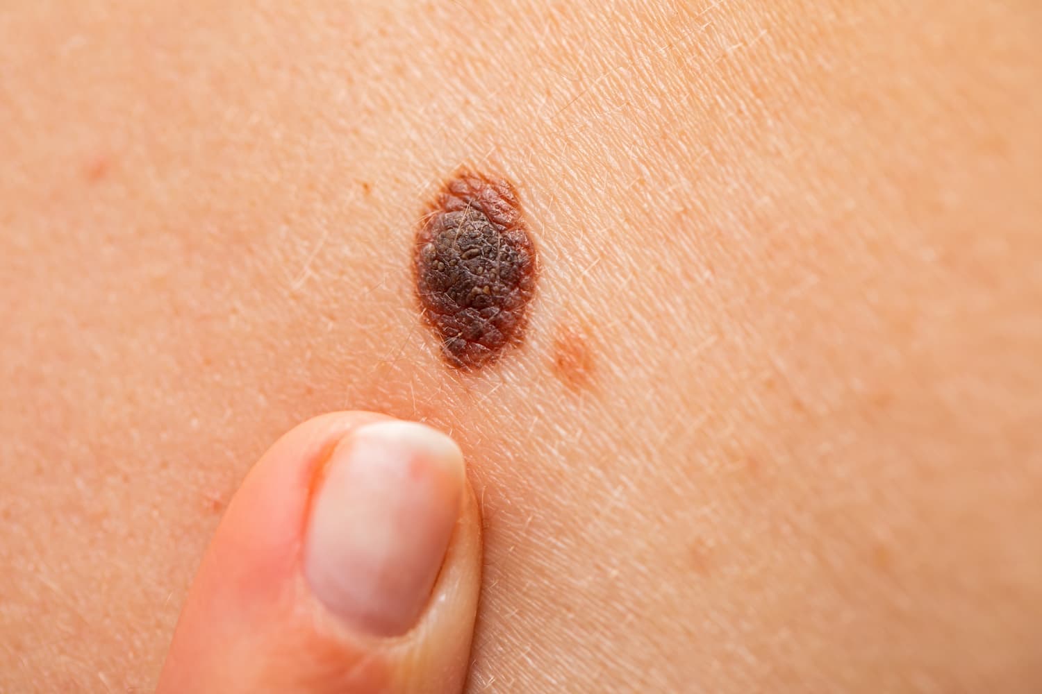
What a mole is
A mole, which doctors call a melanocytic naevus (American spelling: nevus), is a harmless cluster of pigment cells, the melanocytes that give skin its colour (DermNet moles, BAD moles). Almost everyone has at least one, and most adults with light skin have between 10 and 40 (AAD moles overview, NCI). Moles present at birth or appearing within the first two years of life are called congenital; all the others are acquired, and most of those appear in childhood and early adult life (BAD moles, AAD moles overview).
The number of moles keeps rising until about the age of 30 to 40 and then falls, because some moles slowly fade (BAD moles, NCI, MedlinePlus). A mole that appears for the first time in adult life is therefore worth watching, and worth checking if it grows or changes (BAD moles, DermNet moles).
How many moles you have depends on your genes, on how much sun your skin has had and on your immune system. A tendency to many moles runs in families, fair skin and sunburn add more, and treatment that suppresses the immune system increases the number (DermNet moles, BAD moles).
What a benign mole looks like
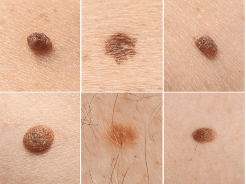
A typical harmless mole is:
- round or oval, with a smooth, even edge (NHS moles, NHS melanoma symptoms);
- one colour throughout, anywhere from pink or skin tone to tan, brown or black; moles are usually darker on brown and black skin (NHS moles, AAD moles symptoms, NCI);
- small, usually no bigger than about 5 to 6 mm, the size of the end of a pencil (NCI, NHS melanoma symptoms);
- flat or raised, smooth or slightly rough, sometimes with a hair growing from it (NHS moles, AAD moles symptoms);
- unchanged from one month to the next (AAD moles symptoms, NHS melanoma symptoms).
Moles can sit anywhere on the skin, including the scalp, between the fingers and toes, on the palms and soles, under a nail, on the genitals and in the eye (AAD moles symptoms, BAD moles). A mole that is flat in childhood often becomes raised and hairier later, and moles on the face tend to lose their colour with age (BAD moles).
Dermatologists name common moles by where the pigment cells sit (BAD moles, DermNet moles):
| Type | What it looks like | When it appears |
|---|---|---|
| Junctional naevus | Flat, usually circular, an even mid- to dark-brown colour | Most childhood moles are this type (BAD moles) |
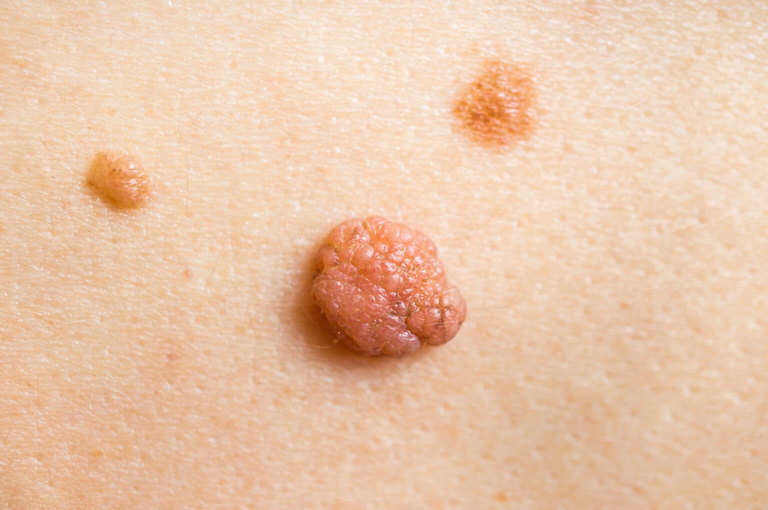
| Compound naevus | A raised brown bump; most are hairy and some have a slightly warty surface | Later in life, as flat moles become raised (BAD moles) |
| Intradermal (dermal) naevus | A raised, often hairy bump that is paler, frequently skin-coloured, sometimes on a small stalk or with a smooth dome | Later still, as a raised mole loses most of its colour; facial moles often pale this way (BAD moles) |
So a raised brown or skin-coloured bump that has been there for years and has not changed is usually a compound or dermal naevus rather than a warning sign (BAD moles). A stable, even, symmetrical mole of this kind is what doctors mean by a benign naevus, whether it is flat or raised, brown or skin-coloured.
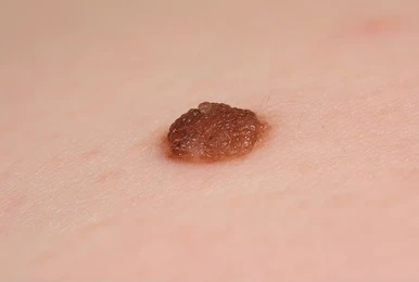
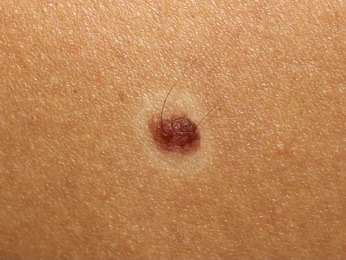
The two photos above show common benign patterns. The first is a raised, evenly light-brown mole with a slightly bumpy surface, consistent with the raised compound or dermal types the BAD describes (BAD moles). The second is a small, flat, evenly brown mole with hairs growing through it; hair in a mole is a normal feature (NHS moles, AAD moles symptoms).
Less common types of mole
| Type | What it looks like | What to know |
|---|---|---|
| Congenital mole (brown birthmark) | Present at birth or soon after; grows in proportion with the child; may darken, thicken and grow hair around puberty (DermNet congenital naevi, NHS birthmarks) | About 1 in 100 babies has a small one, 1 in 1,000 a medium one and 1 in 20,000 a giant one, 20 cm or more in predicted adult size (DermNet congenital naevi). Small and medium congenital moles carry a melanoma risk well under 1 %; large and giant ones carry a real risk and need a dermatologist's follow-up (DermNet congenital naevi, CRUK melanoma risks, NHS birthmarks). |
| Atypical (dysplastic) mole | Larger than 5 mm, oval or irregular, usually flat, sometimes with a raised centre ("fried egg"), a mixture of colours from pink to dark brown, with an edge that fades into the skin (BAD atypical mole syndrome, NCI, DermNet moles) | About 1 in 10 people has at least one (NCI, MedlinePlus). An atypical mole is benign in itself, but it is a marker of higher melanoma risk (see below). |
| Blue naevus | A small, smooth, slate-blue to grey spot, usually 0.5 to 1 cm, on the hands, feet, buttocks, scalp or face; the colour comes from pigment cells lying deep in the skin (DermNet blue naevus) | Common blue naevi stay unchanged for life. One larger than 1 cm, one that changes, or one that appears new in an adult should be checked (DermNet blue naevus). |
| Halo naevus | A mole with a white ring around it; the mole fades over several years and the skin usually regains its colour (DermNet halo naevus, BAD moles) | Mostly children and young adults, about 1 in 100 white-skinned people; linked with vitiligo (DermNet halo naevus). The pale ring burns easily in the sun. In an adult a doctor should check the rest of the skin, because, rarely, haloes appear as a reaction to a melanoma elsewhere (DermNet halo naevus). |
| Spitz naevus | A pink, red-brown or brown dome-shaped bump that grows quickly over a few months, mostly in children and young people (70 % are under 20) (DermNet Spitz naevus, AAD moles types) | It can look like melanoma even to a dermatologist, so it is often removed to be sure (DermNet Spitz naevus, AAD moles types). |
| Meyerson naevus | An ordinary mole surrounded by an itchy ring of eczema (DermNet Meyerson naevus) | The mole in the middle is rarely cancerous, and the eczema settles if the mole is removed (DermNet Meyerson naevus). |
What a dangerous mole looks like: the ABCDE rule
Melanoma is the skin cancer that starts in pigment cells. It can grow inside an existing mole or, more often, on skin that looked normal before (DermNet melanoma, NCI). Estimates of how many melanomas start in a mole differ by source: 20 to 30 % (Skin Cancer Foundation warning signs), 29 % in a 2017 analysis of 38 studies covering over 20,000 melanomas (DermNet melanoma), and about half (HSE moles). Whatever the share, a new dark spot deserves the same attention as a changing old one (HSE moles, NHS melanoma symptoms).
The ABCDE checklist describes the early features of melanoma (DermNet ABCDE, AAD ABCDE, NCI):
| Letter | Melanoma | Benign mole |
|---|---|---|
| A, Asymmetry | One half does not match the other, in shape or in colour | Symmetrical |
| B, Border | Irregular, scalloped, ragged, notched or blurred; the pigment may spread into the surrounding skin (AAD ABCDE, DermNet ABCDE, NCI) | A smooth, even edge; the pigment of a flat mole fades gently towards the edge (DermNet ABCDE) |
| C, Colour | Several shades of brown, black and tan, and sometimes white, grey, red, pink or blue (NCI, AAD ABCDE); half of melanomas include one of these unusual colours (DermNet ABCDE) | One shade, or two arranged evenly (DermNet ABCDE) |
| D, Diameter and Different | Usually more than 6 mm, but smaller when caught early; also the mole that looks different from all your others (NHS melanoma symptoms, DermNet ABCDE) | Under 6 mm and looks like your other moles |
| E, Evolving | Changes in size, shape, colour or height over weeks to months; new itching, bleeding, crusting or soreness (NCI, AAD ABCDE, CRUK melanoma symptoms) | Stable from month to month |
The ugly duckling: Most people's moles resemble each other. A spot that stands out from the rest, whether larger, darker, lighter or simply different, should be checked even if it shows none of the ABCDE features (DermNet ABCDE, CRUK melanoma symptoms, Skin Cancer Foundation warning signs). Fewer than 3 % of all changing skin lesions turn out to be melanoma, and most ugly ducklings turn out to be harmless when a dermatologist examines them with a dermatoscope (DermNet ABCDE, DermNet moles).
EFG for lumps: Nodular melanoma, 10 to 15 % of melanomas, often breaks the ABCDE rules. It is Elevated, Firm and Growing over weeks rather than years, and one in three has no pigment at all (DermNet nodular melanoma, DermNet ABCDE). A new firm lump that keeps growing needs a doctor whatever its colour (DermNet ABCDE).
What melanoma feels like: Most early melanomas cause no pain or itch, so a spot that does not hurt is not automatically safe (Skin Cancer Foundation warning signs, CRUK melanoma symptoms). Itching on its own neither confirms nor rules out melanoma, and some harmless moles itch (Skin Cancer Foundation warning signs, CRUK melanoma symptoms). An itchy mole that is also changing, or that keeps itching, should be checked, and the NHS lists an itchy mole among its reasons to see a GP (NHS melanoma symptoms).
Palms, soles and nails: Melanoma can also start where the sun never reaches. In people with brown or black skin it most often appears on the soles, the palms or under a nail, as a brown or black band running the length of the nail, with pigment spreading onto the skin at the nail fold as a warning clue, or as a dark patch on the sole or palm (NHS melanoma causes, CRUK melanoma symptoms, DermNet acral lentiginous melanoma). A dark line under a nail that was not caused by an injury is a reason to see a doctor, whatever your skin colour (NHS melanoma symptoms, CRUK melanoma symptoms).
Who is more likely to develop melanoma
- Many moles: Risk rises with the count: higher with more than about 50 moles (AAD moles types) and clearly higher with 100 or more compared with 15 or fewer (CRUK melanoma risks, DermNet moles).
- Atypical moles: More than 5 dysplastic naevi roughly multiplies the risk by 10 (NCI); 10 or more by about 12 (Skin Cancer Foundation atypical moles). Atypical mole syndrome, more than 50 moles with at least 3 atypical ones, affects about 2 % of people and carries a 7 to 10 times higher risk; with melanoma in a close relative it is called familial atypical mole syndrome (BAD atypical mole syndrome, CRUK melanoma risks).
- Family history: Any close relative with melanoma raises the risk; it is highest with two or more first-degree relatives or a relative diagnosed under 30 (CRUK melanoma risks, DermNet melanoma). About 1 in 10 melanomas might be linked to an inherited gene change (CRUK melanoma risks).
- Fair skin that burns easily, red or blond hair, blue or green eyes, many freckles (NHS melanoma causes).
- Sunburn and sunbeds: Sunburn at any age raises the risk, and so does intermittent intense sun, for example holiday sun on skin that is usually covered; sunbed use before 35 carries the highest risk (CRUK melanoma risks). Around 85 in 100 melanomas in the UK are caused by too much ultraviolet light (CRUK melanoma risks, BAD melanoma in situ).
- A previous melanoma or other skin cancer, or a weakened immune system, including after an organ transplant (DermNet melanoma, CRUK melanoma risks, NCI).
- A large or giant congenital mole (CRUK melanoma risks, DermNet congenital naevi).
Melanoma is the fifth most common cancer in the UK, with about 18,300 diagnoses a year (BAD melanoma in situ, CRUK melanoma risks). People with black or brown skin get it far less often, with a lifetime risk of about 0.1 % for Black Americans and 0.5 % for Hispanic Americans against 2.9 % for White Americans, but when they do it is often found later and thicker, which is why the nail, palm and sole signs above matter (NCI, DermNet melanoma in skin of colour).
When moles change for harmless reasons
Not every change is a warning. Moles in children grow as the child grows, and some darken while others lighten; these changes are expected and seldom mean melanoma (AAD moles overview). Moles darken after sun exposure and become more raised with age (DermNet ABCDE, BAD moles). As people get older, moles fade or disappear (NHS moles, BAD moles).
Pregnancy: More than 1 in 10 women notice moles darkening or enlarging during pregnancy; moles on the breasts and abdomen commonly enlarge as the skin stretches, while moles on the back and limbs do not change significantly in size (DermNet moles in pregnancy). A slight darkening is normal (NHS moles) and moles usually return to their previous size after the birth (BAD moles). Any ABCDE-type change in pregnancy should still be examined, not put down to hormones (DermNet moles in pregnancy, BAD moles), and a suspicious mole can be removed safely under local anaesthetic during pregnancy (DermNet moles in pregnancy).
Injury and irritation: Raised moles catch on clothing or a razor and can become sore and inflamed after a knock (BAD moles). Bleeding, oozing or crusting remain reasons to have the mole checked, whatever the apparent cause (NHS moles, HSE moles), and any spot that has not healed within about three weeks should be seen (DermNet self skin examination). An itchy ring of eczema around a mole is a Meyerson naevus (DermNet Meyerson naevus).
Pigment in an old scar: After a mole has been shaved or incompletely removed, pigment can reappear in the scar with an odd shape. Doctors call this a recurrent naevus; it should be looked at by the dermatologist who removed the mole, because regrowth can also be a sign of melanoma (DermNet moles, AAD moles treatment).
When to see a doctor
Book a doctor's appointment promptly, which the AAD puts as "immediately", if a mole or dark spot (NHS moles, NHS melanoma symptoms, HSE moles, CRUK melanoma symptoms, AAD moles self-care):
- has changed in size, shape or colour, or has become darker or more raised;
- has two or more colours or an uneven, blurred or jagged edge;
- itches, hurts, is swollen or inflamed, bleeds, oozes or crusts;
- looks different from all your other moles;
- is a new or unusual mark that has not gone after a few weeks, including on the palms or soles; this matters especially if your immune system is weakened (CRUK melanoma symptoms);
- is a dark area or line under a nail that was not caused by an injury.
Also get checked: a mole that appears for the first time after your 30s (HSE moles), and especially after 40 (DermNet moles, Skin Cancer Foundation atypical moles), a birthmark that has become bigger, darker or lumpier, and any large congenital mole (NHS birthmarks).
These changes happen over weeks or months (HSE moles), and any lesion that keeps changing for more than a month should be reviewed by a dermatologist or biopsied (DermNet melanoma). In the UK, a GP who suspects melanoma refers you to a specialist to be seen within 2 weeks (NHS moles); in Ireland the referral is urgent (HSE moles). In the UK, GPs use a weighted 7-point checklist to decide: a change in size, an irregular shape and an irregular colour score 2 points each; a diameter of 7 mm or more, inflammation, oozing and a change in sensation score 1 each; a score of 3 or more triggers an urgent referral, as does a dermoscopic appearance that suggests melanoma (NICE suspected cancer guideline). Moles are not an emergency, but delay does not make the worry go away, and the earlier a melanoma is picked up, the more likely treatment is to succeed (CRUK seeing your GP).
How a dermatologist checks a mole
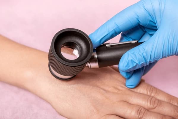
Most moles are recognised by their typical appearance (DermNet moles). The next step is dermoscopy (dermatoscopy): a handheld magnifier with a built-in light that enlarges the mole up to 20 times, is painless and shows the pattern of pigment, the colours, the structure and the border in a way the naked eye cannot (BAD moles, DermNet dermoscopy). In experienced hands dermoscopy can make melanoma easier to diagnose (DermNet dermoscopy). A GP may also measure the mole against a ruler, photograph it, compare it with your other moles and send the pictures to a teledermatology service (CRUK seeing your GP, NHS melanoma symptoms).
For a flat mole that is slightly unusual but not alarming, the dermatologist may repeat the dermoscopic photograph after a short interval to look for change; nodular or suspicious lesions are not monitored this way but removed (DermNet moles, DermNet melanoma). People with many moles or a high risk can have total body photography and digital dermoscopic imaging, often called mole mapping, which records every mole so that new ones and subtle changes show up at the next visit (DermNet moles, DermNet self skin examination).
The only way to know for certain is to remove the tissue and examine it under a microscope (NCI). A suspicious mole is cut out whole, with a 2 mm margin of normal skin, under a local anaesthetic injection; the sample goes to a pathologist (DermNet melanoma, DermNet skin biopsy). Taking only a piece of a suspicious mole is avoided, because a partial sample may miss an area of cancerous change (DermNet moles, DermNet melanoma). A benign mole does not come back after a full excision; a melanoma sometimes does (NCI).
What an online consultation can and cannot do: Clear photographs reviewed by an iDerma dermatologist can give you an opinion on whether a stable mole looks typical, which of your moles deserve in-person dermoscopy and what to watch for. A photograph cannot replace dermoscopy or a tissue sample (NCI, DermNet dermoscopy). A mole with ABCDE changes, a new dark streak under a nail or a growing lump needs hands-on examination; book a mole check with dermoscopy in person for those.
Treatment: when a mole should be removed
Most moles need no treatment and are best left alone (DermNet moles, BAD moles, AAD moles treatment, NHS moles). Removing harmless moles does not lower your risk of melanoma (HSE moles), because most melanomas do not start in an existing mole (NCI). A dermatologist removes a mole for three reasons (DermNet moles, BAD moles, AAD moles treatment):
- there is doubt about the diagnosis, so the mole is cut out and examined to exclude cancer;
- the mole is a nuisance, repeatedly irritated by clothing, a comb or a razor;
- cosmetic reasons; this is not usually provided by the NHS, and in Ireland too it is done privately and can be expensive (NHS moles, HSE moles).
| Method | Used for | What to expect |
|---|---|---|
| Surgical excision | Any flat or suspicious mole, and the standard whenever there is doubt (DermNet moles, AAD moles treatment) | The whole mole is cut out under local anaesthetic and the skin stitched; the mole is examined under the microscope; a fine-line scar is the normal result, and a large mole may need a skin graft (AAD moles treatment, BAD moles) |
| Shave removal | A protruding mole (DermNet moles, AAD moles treatment) | A blade levels the mole with the skin; the sample is usually still examined; pigment can regrow in the scar, which then needs a check (AAD moles treatment, DermNet moles) |
| Electrosurgery and laser | Only once a dermatologist has confirmed the mole is benign; DermNet lists electrosurgical destruction, and laser only to lighten pigment or remove coarse hair (DermNet moles) | No tissue reaches the laboratory, which is why the BAD advises against laser removal of a mole (BAD moles); freezing (cryotherapy) is not used on undiagnosed lesions or on lesions that need pathology (DermNet cryotherapy) |
Every removal leaves some scar (BAD atypical mole syndrome). Darker skin may be left with a temporary dark mark; a pale mark, when it occurs, is usually permanent (DermNet postinflammatory hyperpigmentation). If a removed mole grows back, see the dermatologist again promptly (AAD moles treatment). Read more about mole removal methods and when mole removal is really necessary.
Never remove a mole at home or at a beauty salon: The US Food and Drug Administration warns that products sold for removing moles can cause skin injuries, infections that need antibiotics, scarring and a delayed cancer diagnosis; none is approved (AAD mole removal). If the spot was a melanoma, cells left behind can multiply and spread (AAD mole removal).
Congenital moles: A small congenital mole that grows in step with the child and is not changing in any other way does not have to be removed; if removal is wanted it is usually postponed until the child can cope with a local anaesthetic injection, around 10 to 12 years (DermNet congenital naevi). For a giant congenital mole the decision is individual, partly because half of the melanomas in people with a giant congenital mole arise somewhere other than the mole itself (DermNet congenital naevi).
Prevention and skin care
You cannot prevent moles altogether, but strict sun protection from birth reduces the number, and sunscreen alone is not enough to stop new moles forming (DermNet moles). About a quarter of a lifetime's ultraviolet damage happens before the age of 20 (BAD sun protection for children). The same measures lower the risk of melanoma (NHS moles, HSE moles, CRUK melanoma risks):
- stay in the shade between 11am and 3pm, when the sun is strongest (NHS moles, HSE moles, BAD moles);
- cover up with clothing, a wide-brimmed hat and sunglasses with UV protection (NHS melanoma causes, BAD moles);
- use a sunscreen of at least SPF 30 with 4-star or "UVA" circle protection; the BAD suggests SPF 50 or above for people with a large number of moles (NHS sun safety, HSE moles, BAD moles);
- apply it 15 to 30 minutes before going out, generously (about 6 to 8 teaspoons for the whole body), and again every 2 hours and after swimming or sweating (BAD moles, NHS sun safety, HSE moles);
- never use sunbeds or sunlamps, and do not aim for a tan: a tan is already a sign of damage (NHS moles, HSE moles, BAD moles);
- keep babies under 6 months out of direct sun and protect children with shade and clothing first, sunscreen second (NHS sun safety, BAD sun protection for children);
- start protecting white skin when the UV index reaches 3; the BAD says routine protection is rarely needed for black and brown skin in the UK, with exceptions for a skin condition, a high skin cancer risk, immunosuppression and travel to countries with an extreme UV index (BAD sun protection fact sheet).
Check your own skin: Dermatology bodies recommend a self-check once a month (BAD moles, BAD atypical mole syndrome, NCI, Skin Cancer Foundation warning signs); DermNet suggests every one to three months (DermNet self skin examination). Stand in front of a full-length mirror and look at the front, back and sides with your arms raised; check the forearms, underarms and palms; the backs of the legs, between the toes and the soles; use a hand mirror for the neck, scalp, back and buttocks, and part your hair (AAD skin self-exam, DermNet self skin examination). Ask a partner to check your back, and photograph any mole you are unsure about next to a ruler so that change is easy to see (BAD moles, DermNet congenital naevi).
Professional checks: People with many moles, atypical moles or a previous skin cancer should have regular skin examinations by a doctor (DermNet moles). With more than 5 atypical moles a yearly examination is usual, and every 3 to 6 months if melanoma also runs in the family (NCI). Anyone with 50 or more moles is advised to have a dermatologist who can examine their skin as often as needed (AAD moles self-care), and mole mapping is offered to those at the highest risk (DermNet moles).
Spots that are mistaken for moles
| Looks like a mole | How it differs |
|---|---|
| Seborrhoeic keratosis | A waxy or warty "stuck-on" plaque in any shade from skin colour to black, with a sharply defined edge; appears from the 30s and 40s on, and more than 9 in 10 people over 60 have at least one; not precancerous, but sometimes hard to tell from a skin cancer, in which case it is sampled or removed (DermNet seborrhoeic keratosis, DermNet ABCDE, BAD moles) |
| Solar lentigo (sun spot) or freckle | A flat brown patch with a well-defined edge; solar lentigines routinely evolve over time (DermNet ABCDE) |
| Dermatofibroma | A firm pink to brown nodule, most often on the lower leg, usually 7 to 10 mm, that dimples when pinched; harmless (DermNet dermatofibroma) |
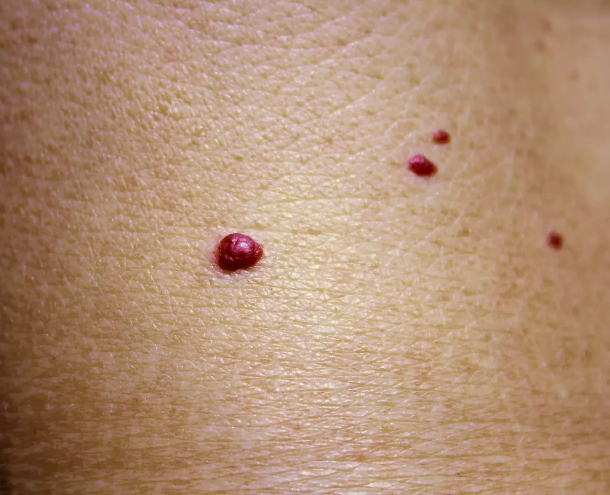
| Cherry angioma | A small red, blue or purple dome, 1 to 10 mm, appearing from about 40; a clotted one can look black until a dermatoscope shows the red (DermNet cherry angioma) |
| Pigmented basal cell carcinoma | A pearly or waxy nodule, sometimes with fine blood vessels or a rolled, ulcerated edge, that can carry pigment and mimic melanoma (DermNet ABCDE, NICE suspected cancer guideline) |
| Lentigo maligna | An early melanoma, not a mole: a slowly growing, flat, freckle-like patch on the sun-damaged face or neck of an older person, enlarging over 5 to 20 years; it is not related to how many moles you have (DermNet lentigo maligna) |
Dermoscopy separates most of these from a mole; when it cannot, the spot is removed and examined (DermNet ABCDE, DermNet seborrhoeic keratosis).
Outlook
Moles that appear in childhood usually stay for life, teenagers and young adults have the most, and the count falls in later life as some fade (DermNet moles). When melanoma is found early, 9 in 10 melanomas can be cured by simple surgery alone, and melanoma in situ, the earliest stage, can be cured if it is completely removed (DermNet melanoma, BAD melanoma in situ). In the United States the five-year survival is 99 % when melanoma is detected early, 74 % once it has reached the lymph nodes and 35 % after distant spread (Skin Cancer Foundation warning signs).
What to do now
- Compare the mole that worries you with the ABCDE table and with your other moles. Photograph it next to a ruler and note the date.
- If any ABCDE sign, an ugly duckling, a new dark streak under a nail or a growing lump is present, see a GP or dermatologist in person promptly.
- If the mole is stable and typical and you want a second opinion or advice on what to watch, book an online consultation: send sharp photos in daylight, close up and from arm's length, with a ruler or coin for scale.
- If you have many moles, atypical moles or a family history of melanoma, set a monthly self-check reminder and ask about a mole check with dermoscopy and mole mapping.