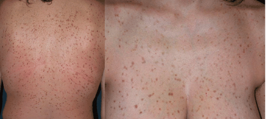
What cutaneous mastocytosis is
Cutaneous mastocytosis is a rare condition in which too many mast cells collect in the skin. Mast cells are immune cells made in the bone marrow; they store histamine and other chemical messengers and release them when they meet something that looks like a threat, which is why a mosquito bite swells and itches (NHS mastocytosis, TMS overview). In mastocytosis a change in a gene called KIT makes the cells multiply and release their chemicals too easily (DermNet mastocytosis). The result on the skin is a set of persistent brown or red spots that redden, swell and itch when they are rubbed.
The word cutaneous means the disease is limited to the skin. When mast cells also build up in the bone marrow, gut, liver, spleen or lymph nodes, the condition is called systemic mastocytosis. The World Health Organization groups mastocytosis into cutaneous, systemic and the very rare mast cell sarcoma (DermNet mastocytosis). Which group a person falls into matters more than anything else for the outlook, and it depends heavily on age: children almost always have the skin-only form, while most adults with the same spots turn out to have some bone-marrow involvement as well (PCDS, Am J Clin Dermatol 2025 review).
Older names you may still read include urticaria pigmentosa and telangiectasia macularis eruptiva perstans (TMEP). Both are now grouped under the term maculopapular cutaneous mastocytosis, the commonest form (BAD urticaria pigmentosa, Am J Clin Dermatol 2025 review). 'Subcutaneous mastocytosis' is not a medical term; people searching for it usually mean the cutaneous form.
Key facts
- Estimates of how common mastocytosis is range from about 1 in 3,500 to 1 in 30,000 people; 1 in 10,000 is the figure most often quoted (NORD, Patient.info, Am J Clin Dermatol 2025 review, PCDS).
- About 90% of skin-limited cases are in children, and 85% of childhood cases appear by the age of two, often in the first months of life (Patient.info, Am J Clin Dermatol 2025 review).
- In children the spots usually fade by puberty. In adults they tend to persist, and only about 1 in 10 adults sees them resolve (NHS mastocytosis, PCDS).
- The spots are not contagious and are not a skin cancer. Indolent (slow) systemic mastocytosis does not shorten life expectancy (NHS mastocytosis, NORD).
- The main risks are severe itch, flushing and, in some people, a severe allergic-type reaction (anaphylaxis). Up to half of adults and up to a fifth of children have at least one such episode over their lifetime, so trigger avoidance and an emergency plan are part of treatment (PCDS, BAD urticaria pigmentosa).
Who gets it
Cutaneous mastocytosis can start at any age, but two peaks stand out. The first is infancy: around half of affected children already have spots before six months of age, and some are born with them (DermNet mastocytosis). The second is early adult life, most often between 20 and 40 (PCDS). Boys and girls, men and women are affected about equally (PCDS, BAD urticaria pigmentosa). Most reported cases are in people with lighter skin; in a large US children's hospital series only 4.7% of children with the condition were Black, against 23% of the clinic's overall patients, although the reasons are not understood (Pediatric Dermatology 2025 cohort).
It does not run in families as a rule. The KIT change happens in the mast cells themselves after conception, so it is not passed from parent to child; a handful of families with more than one affected member have been described (NORD, BAD urticaria pigmentosa).
Causes
Most cases trace back to a change in the KIT gene, which controls how mast cells grow and survive. The change makes the cells more sensitive to a growth signal called stem cell factor, so they multiply and gather in the skin (NHS mastocytosis). One particular variant, called D816V, is found in more than 90% of adults but in only about 40% of children; many children carry different KIT changes, which helps explain why their disease behaves so differently (NORD, Am J Clin Dermatol 2025 review).
The symptoms come from what the extra mast cells release. Histamine widens blood vessels and makes the skin itchy and swollen; other messengers add flushing, stomach cramps and diarrhoea (NHS mastocytosis, NORD). The release can be set off by rubbing the spots, heat, cold, exercise, stress, certain foods and a long list of medicines (see Triggers below).
Nothing you did caused it. There is no diet, product or infection known to bring on mastocytosis, and there is no known way to prevent it (DermNet mastocytosis).
The three types of cutaneous mastocytosis
| Type | Who | What it looks like | Course |
|---|---|---|---|
| Maculopapular cutaneous mastocytosis (urticaria pigmentosa) | The commonest form in children and adults; more than three-quarters start before the age of 10 (BAD urticaria pigmentosa) | Many flat or slightly raised red-brown or tan spots, from 1 mm to several centimetres, mostly on the trunk and thighs. Childhood spots are often larger and more varied; adult-onset spots are small, 3 to 6 mm, and more uniform. Fine visible blood vessels may run through them (NHS symptoms, DermNet mastocytosis). | Childhood form usually fades by adolescence; adult form persists and may slowly spread (DermNet mastocytosis, PCDS). |
| Mastocytoma | Infants, most often diagnosed in the first three months; rare in adults (DermNet mastocytoma) | One to three firm, raised, red, pink, yellow or brown patches or plaques 1 to 5 cm across, sometimes with a pitted orange-peel surface. They itch when rubbed, swell for about 15 minutes afterwards and may blister (DermNet mastocytoma). | Almost all disappear before puberty. Four or more lesions are classed as the maculopapular form (DermNet mastocytoma). |
| Diffuse cutaneous mastocytosis | Rare; starts in infancy, usually the first year (NORD, Patient.info) | Rather than separate spots, the whole skin is red, thickened and leathery, often with an orange-peel texture, intense itch and large blisters that may bleed (DermNet mastocytosis, Patient.info). | The most severe skin form, with a higher chance of flushing episodes, low blood pressure and anaphylaxis; needs specialist care (DermNet mastocytosis, NORD). |
Symptoms: what the spots look and feel like
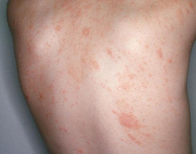
The typical picture is a slowly increasing number of brown or red-brown spots, flat or slightly raised, on the chest, back, tummy and thighs. The face, scalp, palms and soles are usually spared (PCDS, NHS symptoms). The spots are persistent: unlike hives, they do not come and go within hours. People often first notice them as 'new moles' or 'freckles that itch' (BAD urticaria pigmentosa).
The giveaway is Darier's sign: when a spot is rubbed or scratched, it turns red, swells into a hive-like weal and itches within a few minutes, while the skin around it stays calm. Towelling after a bath, friction from clothing and a hot shower are classic triggers (DermNet mastocytosis, PCDS). In young children, particularly under the age of four, a rubbed spot can blister (NORD).
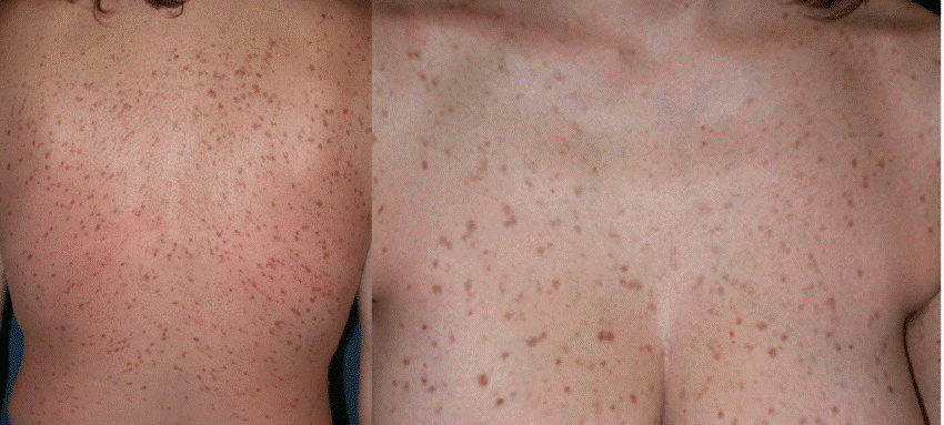
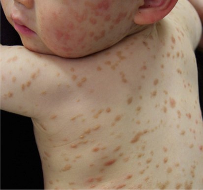
The two photographs above show the maculopapular form in adults (left) and in a toddler (right). The childhood pattern of larger, more varied patches usually fades by adolescence, while the small, uniform adult pattern tends to persist and is the one most associated with systemic disease (DermNet mastocytosis, PCDS, Am J Clin Dermatol 2025 review).
Other symptoms depend on how much histamine the spots release and on whether internal organs are involved (NHS symptoms, Patient.info):
- Itching, which can be intense. Sleep is often affected too: in one study 86% of children with cutaneous mastocytosis scored above the screening threshold for sleep problems, against 31% of healthy children, even when the rash itself bothered them little (Eur J Pediatr 2026 sleep study).
- Flushing of the face or whole body, which affects about half of patients at some point (PCDS).
- A racing or pounding heartbeat, light-headedness or fainting from a drop in blood pressure during a flushing episode (NHS symptoms).
- Stomach cramps, heartburn, nausea or diarrhoea (NORD).
- Headache, tiredness or difficulty concentrating during episodes (NHS symptoms, NORD).
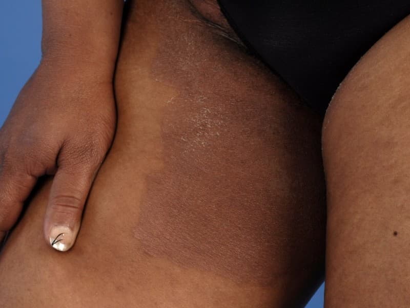
In darker skin the spots can look violet or simply darker than the surrounding skin, and the redness of Darier's sign is harder to see, although the swelling is still there to feel (DermNet mastocytosis). Children with deeper skin tones were more likely to be given treatment in the US cohort; the reasons were not studied (Pediatric Dermatology 2025 cohort).
Cutaneous mastocytosis in adults versus children
The same spots mean different things at different ages.
- Children: the skin is almost always the only organ involved. Blood tests beyond a single tryptase level are not usually needed, a solitary mastocytoma needs no work-up at all, and the spots fade by adolescence in many cases (DermNet mastocytosis, Am J Clin Dermatol 2025 review). Onset after the age of 10 carries a higher chance of the systemic form (BAD urticaria pigmentosa).
- Adults: the vast majority of adults with these spots already have mast cells in the bone marrow, whether or not they feel unwell. Specialists therefore call adult skin lesions 'mastocytosis in the skin' until staging is complete, and a bone-marrow test is considered in every adult-onset case (PCDS, Am J Clin Dermatol 2025 review). About 95% of systemic mastocytosis is diagnosed in adults, and indolent systemic mastocytosis makes up around 90% of adult systemic cases (Patient.info, NHS mastocytosis).
Adult spots also look different: smaller, rounder, darker and more evenly spread, with fewer blisters. This 'monomorphic' pattern is the one most associated with systemic disease (Am J Clin Dermatol 2025 review); the larger, more varied childhood pattern is the one that usually fades by adolescence (DermNet mastocytosis, Patient.info).
Is cutaneous mastocytosis dangerous?
For most people it is not. Skin-limited mastocytosis is benign and does not shorten life, and even indolent systemic mastocytosis has a life expectancy comparable with the general population, with a 3 to 5% risk of progressing to a more advanced form (NORD, Am J Clin Dermatol 2025 review). A Mayo Clinic review of 159 adults found no difference in lifetime survival between those with and without bone-marrow involvement (PCDS). Aggressive systemic mastocytosis and mast cell leukaemia exist but are rare, and there are no reliable markers to predict who is at risk (PCDS).
Three risks need attention:
- Anaphylaxis: Because the body holds far more mast cells than normal, a sting, a medicine or even heat can release a flood of histamine. Up to 50% of adults and up to 20% of children experience at least one episode; in children it is unusual unless the spots are extensive (PCDS, BAD urticaria pigmentosa). Some people learn they have mastocytosis only after an unexplained collapse or a severe reaction to a bee sting (NORD).
- Bone health: Adults with systemic involvement have a higher risk of osteoporosis and may need a bone-density scan (PCDS, NHS mastocytosis).
- Procedures: General anaesthetics, surgery, dental work and X-ray contrast dyes can all trigger mast-cell release, so the team must know the diagnosis in advance (DermNet mastocytosis, BAD urticaria pigmentosa).
Signs of anaphylaxis include a sudden itchy raised rash, swelling of the lips, tongue or throat, wheeze or difficulty breathing, a hoarse voice, feeling faint, cold pale skin, confusion or collapse (NHS anaphylaxis). Use the adrenaline auto-injector if one has been prescribed, call 112 (999 in the UK) immediately, lie the person down with their legs raised (let them sit up slowly if they are struggling to breathe) and do not let them stand or walk; give a second injection after five minutes if there is no improvement (NHS anaphylaxis).
Triggers that can set off the spots
Not every person reacts to every trigger, and triggers can change over time, but the lists from specialist centres overlap closely (DermNet mastocytosis, NHS symptoms, TMS triggers, BAD urticaria pigmentosa):
| Group | Examples |
|---|---|
| Physical | Rubbing, scratching or pressure on the spots; vigorous towelling; tight clothing; hot baths and showers; heat, cold and sudden temperature changes; sunlight; hard exercise; fever |
| Emotional | Stress, anxiety, excitement, lack of sleep |
| Food and drink | Alcohol; spicy food; for some people cheese, shellfish, preservatives and flavourings |
| Stings and bites | Bee and wasp stings above all; jellyfish; biting insects |
| Medicines | Anti-inflammatory painkillers (including some sold without prescription), strong opioid painkillers and cough medicines containing them, some anaesthetic drugs and muscle relaxants used during operations, some antibiotics, X-ray contrast dye, some vaccines. Always remind a prescriber of the diagnosis before starting anything new (BAD urticaria pigmentosa, DermNet mastocytosis) |
| Other | Infections; surgery, dental and endoscopic procedures; strong perfumes and chemical smells (DermNet mastocytosis, TMS triggers, Patient.info) |
Conditions that look similar
A persistent brown spot that swells when rubbed is fairly specific, but several common conditions share one feature or another (DermNet mastocytosis, DermNet mastocytoma):
| Condition | How it differs |
|---|---|
| Hives (acute urticaria) | Weals appear anywhere, last minutes to 24 hours and leave no mark; there is no fixed brown spot underneath (DermNet urticaria overview). |
| Dermographism ('skin writing') | Stroking any area of normal skin raises a line-shaped weal within minutes; it affects about 5% of people and there are no brown spots. In mastocytosis only the spots react (DermNet dermographism, Am J Clin Dermatol 2025 review). |
| Insect bites | Itchy lumps that appear after exposure and settle within days. A mastocytoma is often mistaken for a bite that will not go away (NHS insect bites, DermNet mastocytoma). |
| Moles and brown birthmarks | Stable, do not swell or itch when rubbed. A non-itchy mastocytoma in a baby can look like a congenital mole; a dermoscopy examination and, if needed, a biopsy settle it (DermNet mastocytoma, DermNet congenital naevi). |
| Café-au-lait patches | Flat, light-brown birthmarks with a sharp edge, present at or soon after birth (DermNet café-au-lait macule). |
| Sun spots, freckles and post-inflammatory marks | Flat, uniformly coloured, on sun-exposed skin or at the site of old inflammation; no reaction to rubbing (DermNet solar lentigo, DermNet postinflammatory hyperpigmentation). |
| Cherry angiomas | Small bright red or purple domes that increase in number from the age of 40; they do not itch or swell (DermNet cherry angioma). |
| Seborrhoeic keratoses | Rough, warty, stuck-on brown spots of adult life (DermNet seborrhoeic keratosis). |
| Capillaritis (pigmented purpura) | Flat red-brown patches with 'cayenne pepper' dots, mostly on the lower legs, that do not urticate (DermNet capillaritis). |
| Juvenile xanthogranuloma | A firm yellow-red-brown dome, usually single, on the head or neck of an infant; it fades on its own (DermNet juvenile xanthogranuloma). |
| Langerhans cell histiocytosis | A rare cause of many small pink or red-brown crusted spots on the scalp, body folds and trunk of a young child; needs a biopsy (DermNet Langerhans cell histiocytosis). |
| Bullous impetigo | Thin-roofed blisters that burst into golden crusts; contagious and spreads quickly, unlike the blisters of a rubbed mastocytoma (NHS impetigo, DermNet impetigo). |
| Drug rashes | Appear within hours to days of a new medicine and are usually widespread rather than a few fixed brown spots (DermNet drug eruptions). |
When to see a doctor
- Any brown or red spots that swell, redden or blister when rubbed, in a child or an adult. Confirming the diagnosis early matters because of the trigger and procedure precautions above.
- New spots in an adult: adult-onset disease needs blood tests, and a haematology opinion is considered even if you feel well (PCDS, Am J Clin Dermatol 2025 review).
- Flushing episodes with palpitations, light-headedness, fainting or diarrhoea, with or without visible spots; unexplained anaphylaxis or a severe reaction to a sting should also prompt a mastocytosis check (NORD, Patient.info).
- Bone pain, unexplained weight loss, swollen glands or a swollen tummy in someone with known mastocytosis: these need prompt referral to a haematologist (PCDS).
- Before any operation, anaesthetic, dental procedure or scan with contrast dye, tell the team you have mastocytosis (BAD urticaria pigmentosa).
Call 112 (999 in the UK) for swelling of the lips, tongue or throat, breathing difficulty, wheeze, a hoarse voice, fainting or a child who goes limp or floppy (NHS anaphylaxis).
How cutaneous mastocytosis is diagnosed
Examination and Darier's sign: A dermatologist recognises most cases from the appearance of the spots. The test is simple: a spot is stroked five times with moderate pressure; a positive sign is redness and swelling confined to the spot within a few minutes (NHS mastocytosis, Am J Clin Dermatol 2025 review). The sign is positive in about 90% of maculopapular cases but can be missed in adults, in people taking antihistamine tablets, or if the stroke is too gentle; testing a large nodule too hard in a small child can rarely bring on flushing or wheeze, so it is done with care (PCDS, Am J Clin Dermatol 2025 review).
Skin biopsy: A small punch sample under local anaesthetic shows the crowds of mast cells, which the laboratory highlights with special stains (DermNet mastocytosis, DermNet skin biopsy). In children with a typical picture a biopsy is often unnecessary; in adults, or when the spots could be changing moles, it is usually taken to confirm the diagnosis (BAD urticaria pigmentosa, NHS mastocytosis). The anaesthetic is injected around rather than into the spot to keep the sample readable (PCDS). The biopsy alone cannot say whether the disease is skin-only or systemic (Am J Clin Dermatol 2025 review).
Blood tests: A tryptase level is checked in everyone; tryptase is an enzyme released by mast cells, and a persistently raised level points to systemic disease. Adults also have a full blood count and liver tests. Two tryptase readings of 20 micrograms per litre or more lead to referral to haematology for further investigation (PCDS, DermNet mastocytosis). In children with skin-limited disease no further blood tests are generally needed (Am J Clin Dermatol 2025 review).
Staging tests for suspected systemic disease: These may include an abdominal ultrasound to check the liver and spleen, a bone-density (DEXA) scan, a bone-marrow biopsy taken from the pelvis under local anaesthetic, and genetic testing for the KIT change in blood or marrow (NHS mastocytosis, BAD urticaria pigmentosa). A bone-marrow biopsy is the test that confirms systemic mastocytosis (NHS mastocytosis).
Treatment, by approach
There is no cure for mastocytosis, and skin-limited disease that causes no symptoms needs no treatment at all beyond knowing the triggers (DermNet mastocytosis, NHS mastocytosis). Where treatment is used, it aims to calm the mast cells, relieve itch and prevent severe reactions. Almost all the medicines are used outside their original licence, because trials in this rare condition are scarce (NHS treatment).
- Trigger avoidance is the foundation: lukewarm rather than hot baths, patting rather than rubbing dry, loose clothing, sting avoidance, and a note on your medical record about the medicines to approach with caution (BAD urticaria pigmentosa, DermNet mastocytosis).
- Antihistamine tablets block the main chemical the mast cells release and ease itch, flushing and redness. Modern non-drowsy types are preferred; specialists sometimes prescribe higher than standard doses, and a second type that blocks histamine in the stomach helps acid symptoms (BAD urticaria pigmentosa, DermNet mastocytosis, NHS treatment, PCDS).
- Steroid creams, usually a strong one for a limited time and only on the spots, reduce the number of active mast cells in the skin and improve itch and appearance; long-term use thins the skin, so the risks and benefits are weighed with your dermatologist (NHS treatment, BAD urticaria pigmentosa).
- Mast-cell-stabilising medicines taken by mouth reduce chemical release in the gut and help cramps and diarrhoea; a version applied to the skin exists but is not routinely available (NHS treatment, BAD urticaria pigmentosa).
- Phototherapy with narrowband UVB or light-sensitising medicine plus UVA (PUVA) can reduce itch and fade the spots in more severe cases, although the benefit is often short-lived and the lifetime number of PUVA sessions is limited because of skin-cancer risk (NHS treatment, BAD urticaria pigmentosa, DermNet UVB phototherapy).
- An adrenaline auto-injector is recommended for anyone who has had anaphylaxis, has a high tryptase level or extensive skin disease. UK guidance is that every adult should be offered one, while children with limited spots and no other allergies usually do not need one (DermNet mastocytosis, BAD urticaria pigmentosa). Learn how to use it, carry two, check expiry dates and teach family or carers (NHS anaphylaxis).
- Surgery or laser has a small role: a single mastocytoma in a visible site can be cut out, although the scar may still swell when rubbed, and laser has been used to improve the appearance of adult spots without evidence that the benefit lasts (DermNet mastocytoma, BAD urticaria pigmentosa).
- Medicines for systemic disease are prescribed by haematologists and include targeted tablets that block the faulty KIT signal, injected immune treatments, bone-strengthening medicines and, for the rare aggressive forms, chemotherapy or a stem-cell transplant. Clearing the internal disease can make the skin spots disappear too (NHS treatment, DermNet mastocytosis, NORD).
Medical treatment for the spots themselves is often disappointing. Antihistamines frequently fall short, and many people see only minimal change with phototherapy (PCDS). What treatment reliably achieves is comfort and safety: less itch, better sleep and a plan for a severe reaction.
Living with cutaneous mastocytosis
- Carry the diagnosis with you: wear a medical alert bracelet or carry a card, and tell every new doctor, dentist, anaesthetist or pharmacist 'I have mastocytosis' (BAD urticaria pigmentosa, NHS anaphylaxis).
- Ask about sleep: children with cutaneous mastocytosis sleep worse than their peers even when the rash seems minor, and the study authors recommend asking about sleep at every review (Eur J Pediatr 2026 sleep study).
- Bathing and clothing: use lukewarm water, pat the skin dry, wear soft loose fabrics and avoid heat packs or saunas if they bring on flushing (BAD urticaria pigmentosa).
- Stings: Take simple precautions outdoors in summer and treat any sting as a potential emergency if you have been prescribed an auto-injector (NHS anaphylaxis, PCDS).
- Ask for a joint clinic: Where available, centres where dermatologists, haematologists and allergists work together coordinate long-term review best (BAD urticaria pigmentosa, DermNet mastocytosis).
Outlook
In children the outlook is good. New spots may keep appearing for months or even years before the rash settles, but in many children it has faded by puberty; the younger the child and the fewer the spots, the better the chance of complete clearance (Patient.info, NHS mastocytosis). Mastocytomas almost always vanish before puberty (DermNet mastocytoma).
In adults the spots usually stay, and most adults have some bone-marrow involvement, but the great majority have the indolent form with a normal life expectancy and symptoms that can be managed (PCDS, NORD). Progression to an aggressive form or a blood disorder is rare and is watched for at regular haematology reviews (PCDS, NHS mastocytosis).
What an online consultation can and cannot do
Good photographs of the spots, a short video of Darier's sign after rubbing one of them, and a description of flushing or itch episodes allow an iDerma dermatologist to say whether the picture fits cutaneous mastocytosis, which look-alike conditions can be set aside, and which tests should come next. For a child with typical spots, that assessment plus a tryptase blood test arranged locally is often all that is needed; for an adult, or for anyone with flushing, fainting or stomach symptoms, the dermatologist will direct you to in-person examination, a biopsy and haematology staging. If you have had a sudden severe reaction, do not wait for a consultation: treat it as an emergency.
If you have noticed brown spots that itch or swell when rubbed, or your child has a patch that blisters when touched, book an online consultation. A dermatologist reviews your photographs and history and tells you what the spots are likely to be and what to do next.